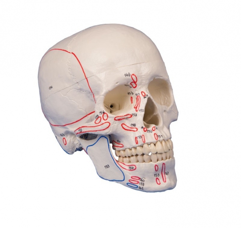
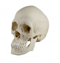
This fascinating natomical model of an average European adult skull can be disassembled into 22 single bones. During development of this model one of the main targets was to make the model easy to assemble and ...
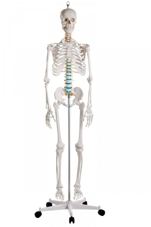
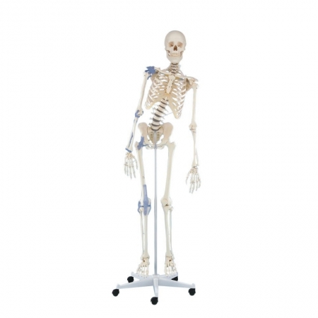
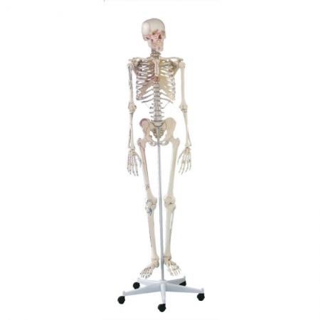
School skeleton. A full-size model of the human skeleton intended for educational purposes. It allows you to demonstrate the anatomical structure of the skeletal system, topography of individual bones and their mutual ...
A flexible model of vertebral column in life size consisting of the occipital plate:
cervical,
thoracic and lumbar vertebrae;
sacrum; coccyx; ]
complete pelvis.
Features include representations ...
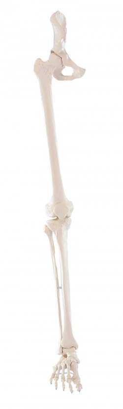
Natural casting of a human leg. Can be dismantled into femur, tibia, fibula and foot. Model with a rubber-mounted foot for increased possibility of movement.
...
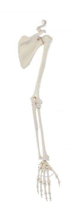
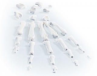
A life-size bone model of the upper limb with a shoulder girdle. Thanks to the flexible connection of the humeral head with the acetabulum, the model allows demonstration of translational movements in the shoulder ...
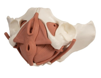
A life-size model of the pelvic floor muscles. Individual muscles can be easily removed. The model also shows the bone anatomy of the female pelvis. The model presents the following structures:
Obturatorius ...
Flexible spine model dedicated to physiotherapists, osteopaths, manual therapists, orthopedists. 1: 1 scale. Possibility to detach the pelvis.
Advantages:
Special assembly on a flexible metal spiral ...
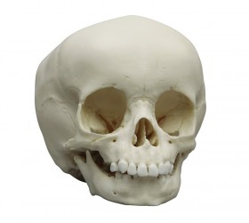
This skull model is a cast of a real human children‘s skull specimen and shows all anatomical structures in great detail. It was developed for students of anatomy, medicine, surgery, ENT medicine, ophthalmology and ...
The human skull with marking of muscle insertions and origins. With nomenclature.
Size: 18 x 19 x 12 cm, weight: 0.7 kg ...
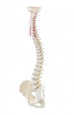
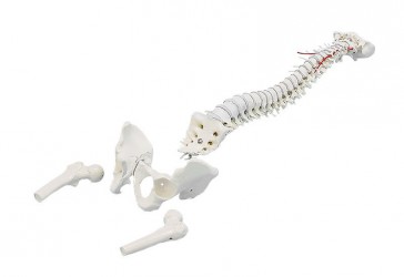
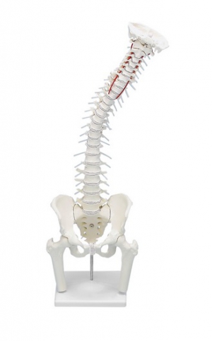
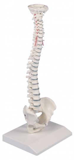
A life-size model of the human spine with a removable pelvis and proximal femoral fragments.
Advantages:
Special assembly on a flexible metal spiral rod that makes the spine stable and at the same time ...
A life-size model of the human spine with a removable pelvis and proximal femoral fragments.
Advantages:
Special assembly on a flexible metal spiral rod that makes the spine stable and at the same time ...
High-quality, natural replica of the human spine with pelvis and femoral bone fragments. A special, flexible tripod enables demonstration of dysfunctions such as:
shortening of the ...
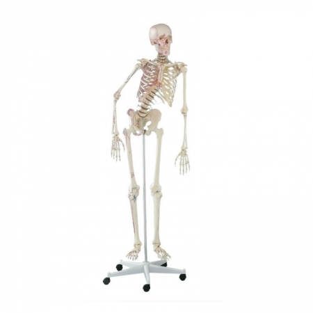
This therapy skeleton with a moveable vertebral column is ideal for anyone who not only wants to learn anatomy, but also wishes as a therapist to understand or explain the connections between movements, postures and ...
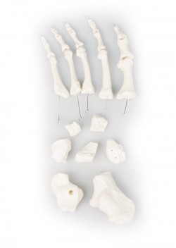
The best quality cast of the adult male's foot bone (life size). ...
The best quality cast of the bones of an adult male. ...
"Pocket" model of the spine. Perfect for seminars, trips or to the office.
Spine reduced by about half its natural size. All dice are shown separately. Movable model. ...
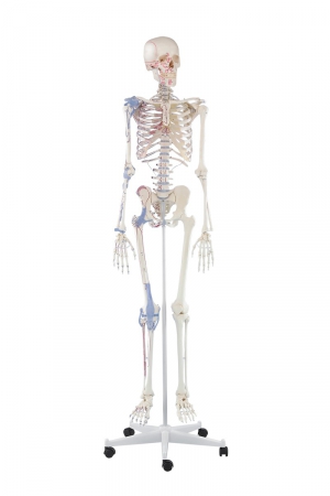
This therapy skeleton offers articular ligaments on one body side in addition to the moveable vertebral column.
The model has the following characteristics:
Natural casting of a human skeleton
...
Anatomical skeleton with muscle marking. All anatomical structures and details are represented in detail.
The model has the following characteristics:
Natural casting of a human skeleton
Representation of ...
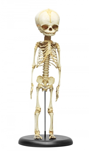
A detailed skeleton of a human fetus on the base.
Average body length measurements of this skeleton suggest an age of 8.5 to 9 months, but developmental (non-metric) osteological traits are most suggestive from 7 to ...
This therapy skeleton offers marking of the muscle origins and insertion points on one body side in addition to the moveable vertebral column.
The model has the following characteristics:
Natural casting of ...
Anatomical skeleton with articular ligaments as well as muscle marking. All anatomical structures and details are represented in detail.
The model has the following characteristics:
Natural casting of a human ...
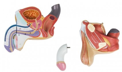
This life size 4 part model is dissected through the median sagittal plane and provides excellent views of external and internal structures. The removable parts include two halves of the penis, showing medial and ...
An excellent example of a condition in which the skull is abnormally long and narrow, as a result of premature closure of the sagittal suture, with heavy centers of ossification in the line of the suture.
Size: 20 x ...
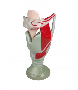
Enlarged Model of a human larynx with hyoid bone. It shows cartilaginous structures on the right side, the left side shows musculature. Movable mounted are vocal cords, arytenoid cartilages and epiglottis to ...
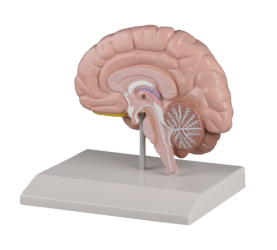
A life-size model of the right cerebral hemisphere with the cerebellum and brainstem shown.
specifications:
dimensions: 16cm x 13cm x 18cm
weight: 0.3 kg
advanced painting
delivered based ...
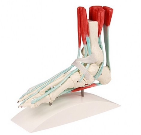
Life size foot skeleton with representation of the ligamentous apparatus with related muscles. All bones of the foot as well as start of tibia and fibula are represented separately. The following muscles with ...
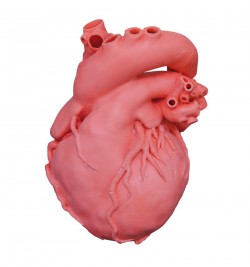
The pediatric congenital heart disease model is based on actual CT data, and is a precisely produced, urethane based, soft model. This product was created with the intention of being a support tool for doctors ...
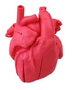
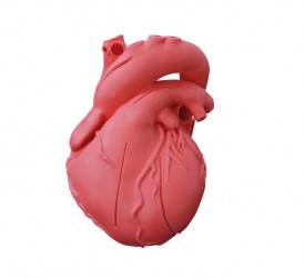
This 2-times life size heart model is based on CT scan data of a healthy, adult male and is anatomically correct inside and out. Reproduced sections: (external & luminal surfaces) right atrium, left atrium / ...
2-times life size heart model is based on CT scan data of a healthy, adult male and is anatomically correct inside and out. Reproduced sections: (external & luminal surfaces) right atrium, left atrium / right ...
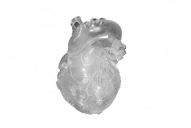
This world-wide unique model is based on CT scan data of a healthy, adult male and is anatomically correct inside and outside. The heart is made of soft and lifelike material. It is pre-cut at different positions to ...
Natural casting of a human leg. Can be dismantled into femur, tibia, fibula and foot. With removable half pelvis.
...
First class actual cast of a 15 months old child in extraordinary high detail.
Due to the very special production technology even smallest details are reproduced and the model looks and feels almost ...
Anatomical Models
Anatomical models for the anatomy laboratory - teaching aids for learning anatomy
Anatomical models are all kinds of teaching aids, presenting the structure and function of individual organs, organs and body systems. They facilitate learning by accelerating the learning process, which is possible by detailing the individual elements of a given body part and understanding its physiology.
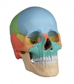
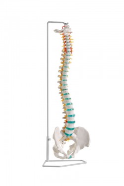
Some of them are natural casts of human bones and organs. They are perfect equipment for anatomical, biological and natural science laboratories. They are ideal as teaching aids for medical students, doctors, manual therapists and osteopaths. Particularly noteworthy is the high-class osteopathic skull, consisting of 22 separate elements, connected by discreetly hidden magnets. It is a model available in two versions - didactic (color) and anatomical (white). A wide range of anatomical skeletons allows you to choose the appropriate model depending on the educational needs of a given group of recipients. The Oscar skeleton is the basic version of a didactic model designed to study the anatomy of the human skeletal system. Anatomical skeletons also have additional accessories, e.g. flexible spine (Hugo's skeleton), joint ligaments (Otto's skeleton), markings of muscle attachments (Arnold skeleton). There is also a version that combines all these elements in one - MAX Skeleton. The flexible model of the spine, with individual vertebrae and intervertebral discs mounted on an elastic spiral rod, is highly mobile. It significantly facilitates the study of anatomy, biomechanics and the study and mobilization of the spine joints in two- and three-dimensional locking.
The anatomical models in our offer:
- Human Skeleton Model: Oscar Skeleton, Hugo Skeleton, Bert Skeleton, Arnold Skeleton, Otto Skeleton, MAX Skeleton.
- Spine model: basic, advanced, with fragments of the femurs,
- flexible skull model (e.g. educational osteopathic skull, divided into 22 parts),
- anatomical models of joints and skeletons models of the upper and lower limbs anatomical models of organs, systems and organs
- Veterinary Models: Cow Model, Sheep Model, Pig Model, Horse Model, Organ Models and Animal Skeletons.
The anatomical models in the form of 3D prints - New!
High-fidelity anatomical models made in 3D printing technology. Three-dimensional anatomical prints are high-quality exhibits based on radiological data and the use of 3D printers, thanks to which they reflect real human bodies. The use of 3D printing technology is an innovative solution in the field of anatomy science, gaining more and more interest among Polish and foreign medical universities. The groundbreaking series of three-dimensional anatomical prints (Monash 3D Printed Anatomy Series) is a unique and one-of-a-kind collection of human anatomy exhibits, designed with teaching in mind, in order to improve the standards of anatomy learning and provide safe and hygienic working conditions for academic teachers and medical students.


 See our profile on Facebook
 Check our profile on Instagram
 Download a PDF file
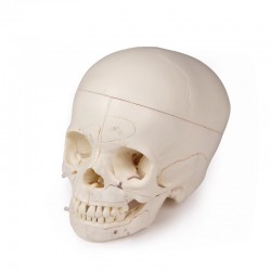


