Quality Certyficate
street: Kolejowa 2, 30-805 Cracow


Home / Anatomical Models / Digestive system models
Anatomical Models
3D anatomy models
Custom tools for patient education
Veterinary simulators
Anatomical Charts
Anatomical Table 3D
Medical simulators
Medical Equipment
Type:
 See our profile on Facebook
 Check our profile on Instagram
 Download a PDF file
Pancreas model

Digestive system model
Digestive system model - miscellaneous
Liver model
Stomach model
Anatomical Models / Digestive system models
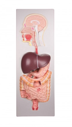
This life size model shows the human digestive tract from mouth cavity to rectum. The oral cavity, the pharynx and the first part of the esophagus are dissected along the medial sagittal plane. The liver is shown ...
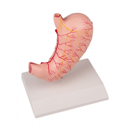
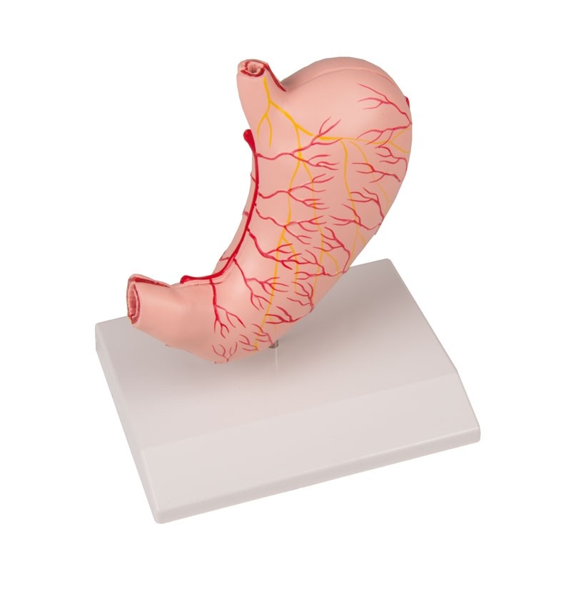
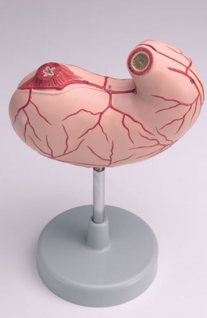
This life size model is dissected along the medial plane and can be opened to show the internal structure of the stomach, including the cardia, the mucosa and the pylorus. The model also shows the blood vessels. ...

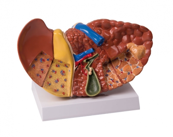
The organs are shown partially opened, and the gall bladder is removable with part of the liver.
It depicts:
Pancreas
Duodenum
Gall bladder
Spleen
Kidneys
Adrenal glands
Blood ...

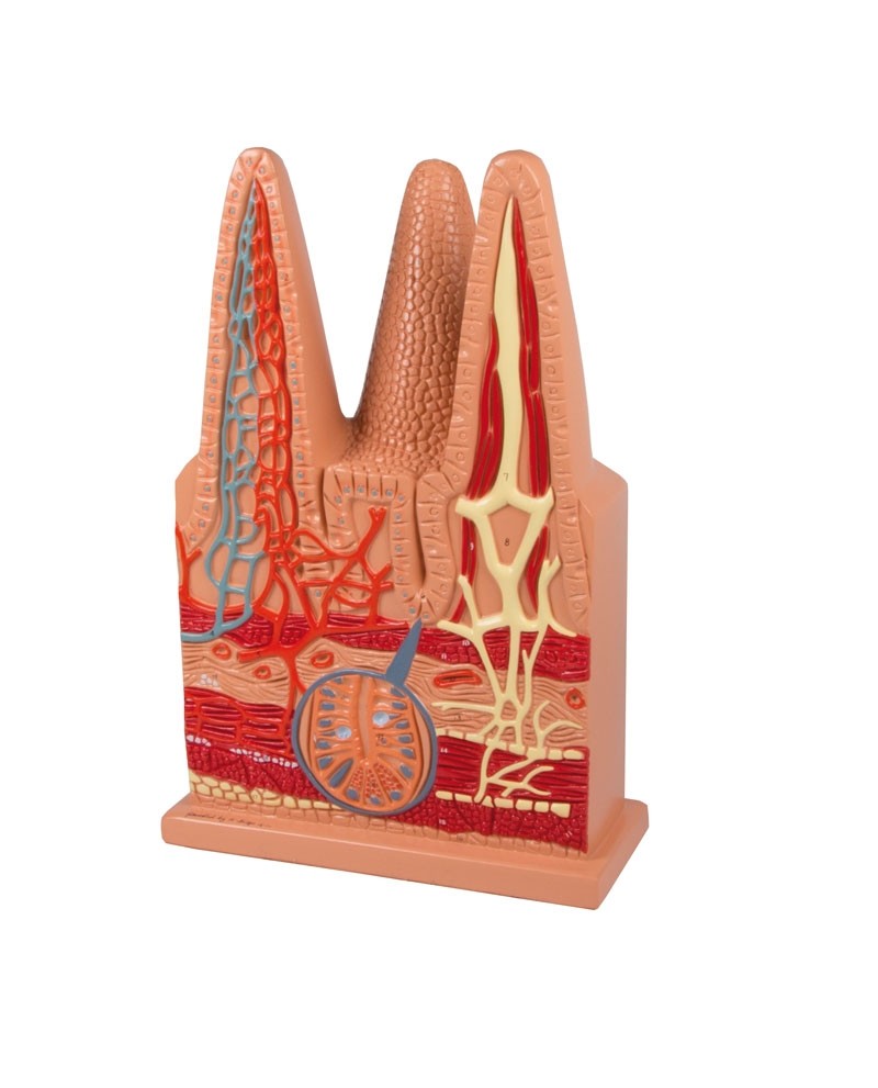
This 100 times life-size model consists of one entire villus, one longitudinally sectioned villus showing the arterioles and venules and one cross-sectioned villus to show the lymphatic vessels. Also includes a ...
This life size model is dissected along the medial plane and can be opened to show the internal structure of the stomach, including the mucosa, the pylorus, and a section of the gastric wall. The model also shows the ...
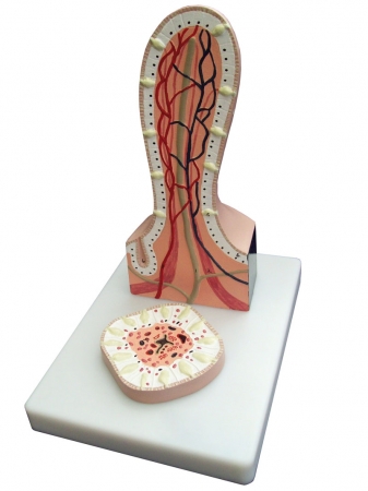
This greatly enlarged 2-part model of a villus from the small intestine shows detail from a transverse and longitudinal section. Mounted on base, with key card.
Size: 17.5 x 15 cm ...
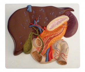
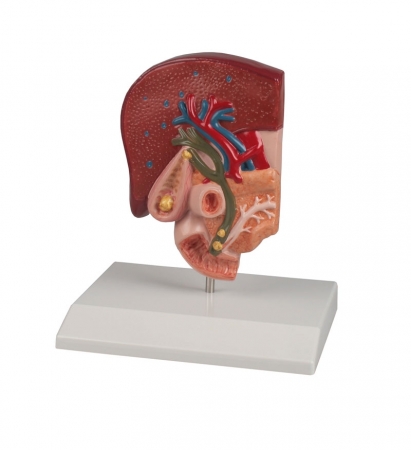
This life size model shows a section of the liver with gall bladder, pancreas and duodenum; includes hepatic and pancreatic ducts. Mounted on board.
Size: 4 x 20 x 18 cm ...
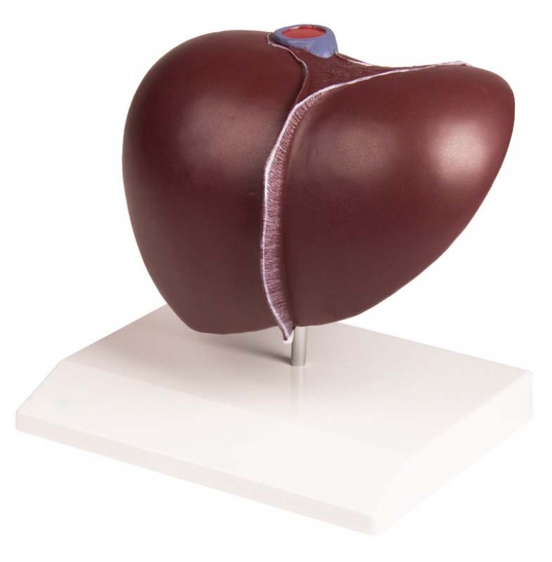
Full size liver model shows conditions such as: Cirrhosis (septal and nodular), biliary obstruction, gall stones, and tumors.
Size: 20 x 11.5 x 14 cm ...
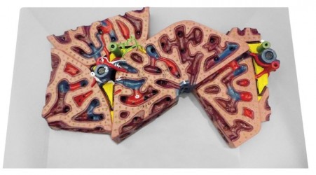
This greatly enlarged model shows the fine detail of a single liver lobule, which is sectioned and shown in relationship to portions of surrounding lobules. The fine colouring distinguishes the portal veins and ...
This half natural size model shows the anatomy of the biliary system and ist surroundings in great detail. Both the tissue changes caused by chronic inflammation and acute inflammation (cholecystitis) are represented ...
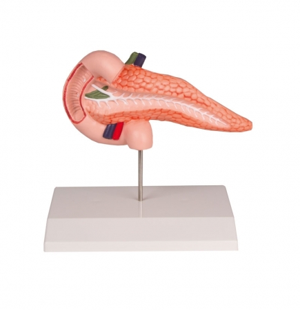
This life size model is an accurate representation of the pancreas and duodenum. The pancreas is open to show the entire pancreatic duct. The duodenum is partially dissected to expose its internal structure. Mounted ...
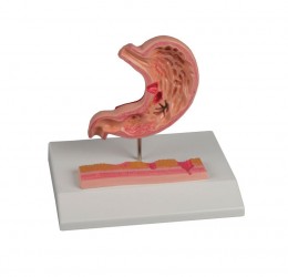
This reduced size model shows different stages of gastritis starting from light ulcer and ending in perforation. The section model shows the lower part of esophagus, the stomach and the start of duodenum.
It shows ...
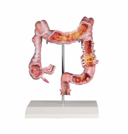
Reduced size model of a human colon showing ileum, caecum, ascending colon, transverse colon, descending colon, sigmoid colon and rectum. The following diseases are depicted: appendicitis, Crohn‘s disease, ...
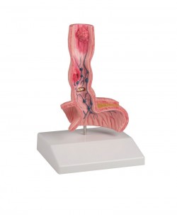
This life-size model shows a frontal section of the lower part of the oesophagus and the upper part of the stomach.
The most common diseases are depicted:
Hiatal hernia
Ulcer
Reflux oesophagitis
...
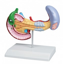
This full size model shows pancreatic cancer, the gallbladder with stones, a ruptured spleen and duodenum with an ulcer. ...
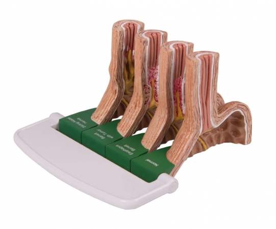
This 4-piece model shows the progressive stages of GERD.
The conditions include:
Sliding Hiatal Hernia and Acid Reflux; Chronic Acid Reflux/Barrett‘s Esophagus; Barrett‘s Esophagus/Adeno Carcinoma.
Size of single ...
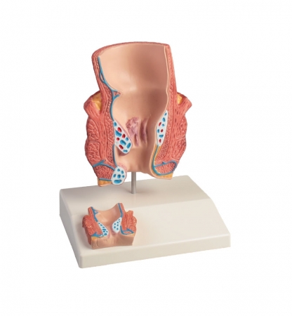
This descriptive model for patient education in about twice life size shows a frontalsection through the rectum. Additionally a smaller section can be found on the base as relief. The model shows outer haemorrhoids ...
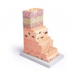
This one-piece model, 150 times life size, is an important tool to study the histology of the most important organ of the digestive system: the stomach. All the different layers from the epithelium to the serous coat ...
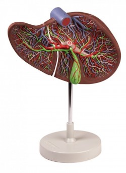
Enlarged 1.5 times, this model shows a liver that is dissected to expose the internal distribution of arteries and veins, the portal vein and the bile duct. Mounted on stand.
Size: 15 x 26 x 12 cm, Weight: approx. 1 ...
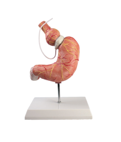
This life-size stomach model shows the function of the gastric band. This model clearly shows the effect of this band used in morbid obesity. The model can be split in two halves to show internal anatomical ...
1
Anatomical Models - Digestive system models
Models of the digestive system are irreplaceable educational tools and practical elements of equipment for medical and scientific offices. These models are used mainly in primary schools, high schools and medical universities. Medical specialists such as doctors, physiotherapists and osteopaths equip their offices with them to serve a didactic and decorative function. Anatomical models offer visual and three-dimensional representations of various parts of the digestive system, making it much easier to understand its complex structure and functions. In our online store, OpenMedis, we have a wide selection of anatomical models of the digestive system that can be used for educational, research and practical purposes.
Anatomy of the digestive system:
The digestive system is a complex system of organs and structures that plays a major role in digestion, absorption of nutrients, and excretion of waste.
The digestive tract is a long tract that begins with the mouth and leads through the throat, esophagus, stomach, small intestine and large intestine. The processes of digestion and absorption of nutrients take place here.
- The stomach - is an organ responsible for the temporary storage and digestion of food. It secretes digestive juices that help break down food into more digestible forms.
- Liver - is the largest organ of the digestive system. This organ performs many different functions, including producing bile, detoxifying the body, and storing glucose.
- Pancreas - helps break down nutrients from food and produces hormones such as insulin, which regulates blood sugar levels.
- Small Intestine - This is the site of the main absorption of nutrients that have been digested in earlier stages.
- Large intestine - responsible for water absorption and the process of stool formation.
The digestive process in the digestive system involves various nutrients that are digested at different stages.
- Proteins - Protein digestion begins in the stomach with protease enzymes such as pepsin, which break them down into shorter polypeptide chains.
- Carbohydrates - their digestion takes place in two stages. Salivary amylase, which is secreted in the mouth, breaks down carbohydrates into shorter sugars. In the intestines, the pancreas produces pancreatic amylase, converting carbohydrates into simpler sugars such as glucose.
- Fats - the first, initial stage of fat digestion begins in the stomach with the help of gastric lipases. The main one takes place in the duodenum, where pancreatic lipase converts fats into glycerol and fatty acids.
- Fiber - fiber is not digested in the human digestive system, but it has a positive effect on the health of the digestive tract and regulates bowel movements.
- Vitamins and Minerals - are absorbed mainly in the small intestine.
- Water - is absorbed along the entire length of the digestive tract, but especially in the intestines, where most nutrients are absorbed.
What does the OpenMedis online store offer?
Due to the wealth of various anatomical structures and the complicated anatomical structure of the human digestive system, anatomical models are helpful in education in this area. Our online offer includes a large variety of models of the human digestive system.
- Digestive system models - we have life-size models of the digestive system in our resources. They present structures from the mouth to the anus. Some elements are shown in the sagittal plane. Additionally, in the case of some internal organs, we can install them.
- Stomach models - our online store has a wide range of stomach models. We can offer you full models with removable elements, cross-sectional models of the stomach, enlarged models of the stomach wall and models of the stomach after surgery for obesity.
- Liver models - mainly present the liver with gallbladder. Our offer also includes comprehensive models of the liver with pancreas and duodenum. For lovers of microanatomy, we present an enlarged model of a liver lobe.
- Pancreas models - our offer includes models of the pancreas combined with the duodenum and models of pancreatic diseases.
Application of anatomical models of the digestive system:
Our anatomical models of the digestive system are an excellent tool for learning and demonstrating various aspects of the anatomy and physiology of this system. Here are some applications of these models:
- Education of students of medicine, biology and other fields related to natural sciences.
- Doctors and other specialists use these models to explain to patients the conditions, diagnostic procedures and treatments related to their digestive system.
- Anatomical models of the human digestive system make it easier to explain complex issues related to the digestive system during presentations and lectures.
The anatomical models of the digestive system available in our online store are made of high-quality materials, which ensures their durability and accuracy in reproducing anatomical structures. Whether you are a student, teacher, doctor or researcher, our models will help you gain the knowledge and skills you need to understand and work with the digestive system.
We also recommend skull models and human torso models, which, combined with anatomical models of the digestive system, will create an ideal set for learning anatomy.
If you have questions or need a specific model of the digestive system, please contact us by e-mail or phone. We are ready to help and provide you with the necessary materials for your study and research work.
We can produce models to order.
