Quality Certyficate
street: Kolejowa 2, 30-805 Cracow


Home / 3D anatomy models
3D anatomy models
Anatomical Models
Custom tools for patient education
Veterinary simulators
Anatomical Charts
Anatomical Table 3D
Medical simulators
Medical Equipment
Type:
 See our profile on Facebook
 Check our profile on Instagram
 Download a PDF file
Our bestsellers
3D anatomy models
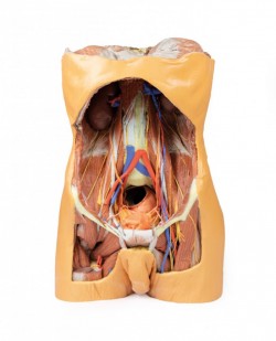
This large, multipart 3D printed specimen displays the entire male posterior abdominal wall from the diaphragm to the pelvic brim, as well as pelvic anatomy and to the proximal thigh. This same individual specimen is ...
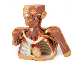
This large, multipart 3D printed specimen displays a great deal of anatomy spanning the head, neck, thorax, axillae and upper limbs. Head and neck: The head and neck of the specimen provides views of both ...
This 3D brain stem anatomical model preserves the several deep cerebral and diencephalic structures through to the proximal medulla oblongata and compliment the other isolated brainstem (BRW10) in our series. ...
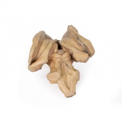
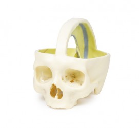
This unique 3D model has been created from CT imaging and segmentation of the internal spaces of the viscerocranium. Parts of the skull have been retained but sections or windows have been removed to expose the ...
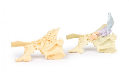
This 3D model preserves a transverse section through the cranial cavity with partial dissection of the brain and exposure of the left orbital roof, alonside a deep dissection of the face and temporomandibular joint ...
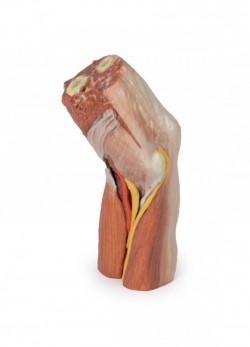
This 3D printed specimen presents a left distal arm and proximal forearm with all skin, subcutaneous fat and superficial cutaneous nerves and veins removed. The elbow region partially flexed to display the arrangement ...
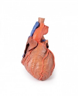
This 3D printed specimen preserves the external anatomy of the heart and the distal trachea, carina, and primary bronchi in the posterior mediastinum relative to the great vessels and left atrium (which demonstrates the ...
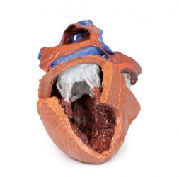
This heart model has been dissected to display the internal structures of the chambers. At the base of the heart the termination of the superior vena cava is preserved entering the right atrium. Part of the inferior ...
Clinical History
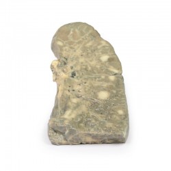
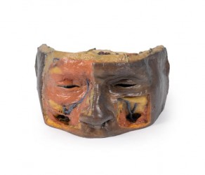
This 47 year old woman was admitted with terminal carcinomatosis. On examination a hard liver and a right pelvic mass were palpable. She had been ill with constitutional symptoms for months and she ...
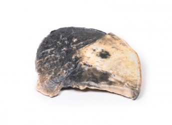
Pathology
The specimen is a parasagittal section of the right lung and the boundaries between the three lobes are visible. The entire upper and middle lobes are congested and hyperaemic* causing the darker appearance. ...
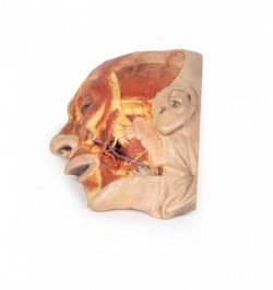
This 3D model presents a superficial dissection of a left face anterior to the ear with false colouring highlighting a series of neurovascular structures alongside the superficial muscles of facial expression. This ...

This 3D model provides a view of the isolated brainstem anatomy from the midbrain to the medulla oblongata, and compliments the other diencephalon/brainstem 3D model (BR 10) in our series. Rostrally, the 3D model has ...
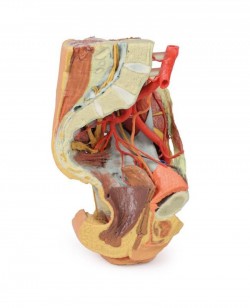
This 3D printed female left pelvis and proximal thigh preserves both superficial and deep structures of the true and false pelves, inguinal region, femoral triangle, and gluteal region. The specimen has been sectioned ...
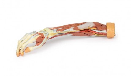
This 3D print of a superficially dissected right upper limb specimen displays a mixture of the vascular, nervous and muscular anatomy of the distal arm, forearm and hand. In the distal arm and elbow/cubital fossa ...

This 3D printed specimen compliments our dorsal dissection specimen (AM01273) by presenting a ventral deep dissection of axial anatomy from the head, neck, axillae, thorax, and abdomen to the proximal portion of the ...
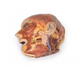
This 3D model presents the superficial anatomy of the face and head, and compliments the superficial facial anatomy of our HW 44 model with a more expanded dissection across the scalp and occipital regions. The ...
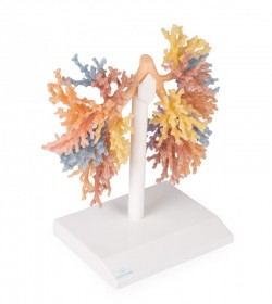
The highly detailed 3D model of the bronchial tree shows the airways from the trachea, spurs trachea and the entire right and left bronchial tree to the bronchial level of the tertiary lobes. Each lobe bronchus system ...
This 3D printed model captures a dissection in which the calvaria and cerebrum have been removed to expose the floors of the anterior and middle cranial fossae. The midbrain has been sectioned at the level of the ...
This 3D printed specimen shows the orbit from the lateral perspective when the bony lateral wall and part of the calvaria of the skull have been removed. The frontal and temporal lobes of the brain are exposed. In ...
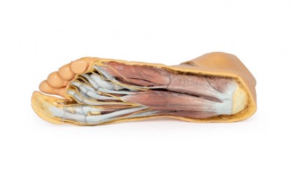
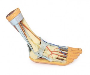
This 3D print records the anatomy of a right distal leg and the deep structures of the plantar surface of the foot. Proximally, the tibia, fibula, interosseous membrane, and leg muscles are discernable in ...
This 3D printed specimen preserves a dissection of the right thoracic wall, axilla, and the root of the neck. The specimen is cut just parasagittally and the visceral contents of the chest have been removed. Structures ...
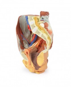
This 3D printed male left pelvis and proximal thigh (sectioned through the midsagittal plane in the midline and transversely through the L3/4 intervertebral disc) shows superficial and deep structures of the true and ...
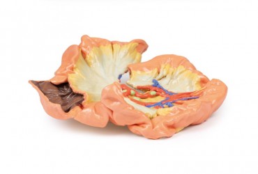
This 3D printed specimen presents a small loop of jejenum and mesentery. A window into the mesentery, fat and visceral peritoneum has been removed to illustrate the arterial arcades in the mesentery (many long ...
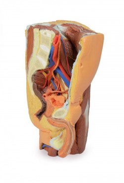
This 3D printed specimen represents a female right pelvis, sectioned along the midsagittal plane and transversely across the level of the L4 vertebrae and the proximal thigh. The specimen has been dissected to ...
This 3D printed specimen presents a deep dissection of a left pelvis and thigh to show the course of the femoral artery and sciatic nerve from their proximal origins to the midshaft of the femur. Proximally, the pelvis ...
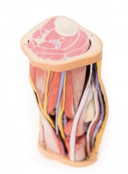
This 3D printed specimen preserves the distal thigh and proximal leg, dissected posteriorly to demonstrate the contents of the popliteal fossa and surrounding region. The proximal cross-section demonstrates the ...

This 3D printed specimen provides a view of deep plantar structures of a right foot. Medially, the cut edge of the great saphenous vein is visible within the superficial fascia, just anterior to the cut edges of the ...
This 3D print of a dissected and opened cranial cavity displays the dural folds and dural venous sinuses, including the falx cerebri (preserved by a retained midsagittal portion of the calvaria. The intact tentorium ...
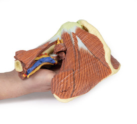
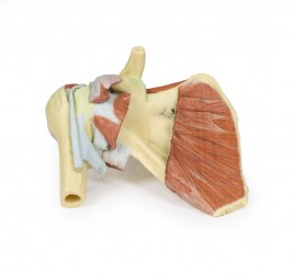
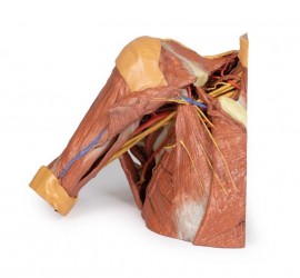
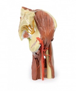
This 3D printed specimen presents a deep dissection of the left shoulder joint, musculature, and associated nerves and vessels of the scapula and proximal humerus (to near midshaft). Anteriorly, the deltoid muscle ...
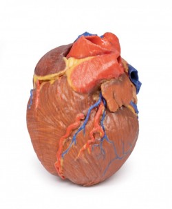
This 3D printed heart specimen preserves superficial cardiac anatomy and the bases of the great vessels. All four chambers (atria and ventricles) are preserved, with the pericardial reflections on the left atrium ...
This 3D printed specimen presents a small loop of jejenum and mesentery. A window into the mesentery, fat and visceral peritoneum has been removed to illustrate the arterial arcades in the mesentery (many long ...
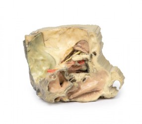
This 3D print displays the orbital contents and its close relations as viewed from the medial perspective when the majority of the lateral wall of the nasal cavity and the intervening ethmoidal sinuses have been ...
3D anatomy models
High Fidelity Anatomical Models - Application
3D anatomical models are very high-quality, detailed and very precisely made teaching aids for learning anatomy, life-size. They present the anatomical parts of the human body - muscles, nerves, blood vessels, internal organs in the so-called high fidelity. This means that individual structures are presented in these models in precise anatomical courses and topographic positions, and the appearance of these structures presents the actual state in a very similar way as in the real human body. It is possible thanks to the combination of production methods consisting in graphic processing of images generated from CT and innovative 3D printing technology. Thanks to these modern models, students have the ability to precisely learn human anatomy, just like on real prosectorial preparations. They can get acquainted with the details of the human anatomy - head, limbs, torso, organs and other structures of the human body. The use of unconventional anatomical models in the form of 3D prints is a pioneering and modern solution in the field of anatomy science, which is becoming more and more popular among Polish and foreign Medical Universities. The breakthrough series of 3D anatomical models is a unique and one-of-a-kind collection of human anatomy exhibits, designed with teaching in mind, with the aim of improving the standards of anatomy learning and providing equal, safe and hygienic working conditions for academic teachers and medical students. 3D anatomical models also have another important advantage - they do not wear out over time, so that each student is provided with the same, identical conditions for learning. The use of detailed anatomical models in the form of 3D printouts in classrooms enables classes in small groups using the latest educational materials and technological solutions. Using such models, students can not only learn the detailed anatomical structure of the human body, but also practice performing a specific type of surgery - plan how to prepare well for it and how to perform it precisely. The modern equipment of the anatomical laboratory makes it easier for students to learn such an important subject as anatomy, and academic teachers conduct classes at a high didactic level, thanks to which their students will be able to achieve the assumed educational results more easily and faster.
Why choose 3D Anatomical Models?
Thanks to 3D printing technology, more and more high-fidelity anatomical models are appearing on the market. They are characterized, above all, by the extraordinary precision of workmanship. In addition, these models are made in a 1: 1 scale or they can also be produced in an enlargement or reduction according to the needs of a given user. The undoubted advantage of 3D anatomical models is their low wear and tear, so they can be used many times in classrooms and provide equal teaching opportunities for students and lecturers. Contrary to prosectoring preparations, 3D anatomical models do not require special storage and disposal conditions, which significantly reduces operating costs. They can be used in ordinary classrooms (they do not require conducting classes in dissecting rooms and laboratories equipped with specialized equipment). Another positive aspect of choosing 3D anatomical models as equipment for your studio is the fact that they are made entirely of artificial materials, so they certainly ensure occupational health and safety for both students and academic teachers. Overall, 3D anatomical models are an excellent alternative to plastinates. In addition, access to them is much faster, cheaper and easier than in the case of cadaver preparations. By comparison, the currently used educational tools in the form of "human bodies" or plastinates have many shortcomings. For example, these are the high costs of preparation, For For Comparing the currently used educational tools in the form of "human bodies" or plastinates with models in the form of 3D prints, one can find many shortcomings. For example, there are high costs related to preparation, storage and disposal. Moreover, the very process of their production is very time-consuming. Prosectoral preparations require special conditions in which they can be used, and therefore they are not available for every group of users (wet preparations cannot be used outside mortuary). Kinds of high fidelity models The only limitations in this area are the imagination and, in extreme cases, the scale of the project. The size and accuracy of such a model depends on the needs of a given project. We can print everything from the smallest particles of organisms to individual internal and external organs, up to comprehensive human or animal models.
What high-fidelity anatomical models does OpenMedis offer?
The range of our products is practically unlimited.
Thanks to the cooperation with us, we are able to meet the expectations of the most demanding customers. Our assortment includes both complete and smaller models of specific organs or body parts. These are largely models made in response to market demand. These are among others:
- Human body with nervous system and organs
- Cross-section of the female pelvis, superficial and deep structures
- Muscles of the dorsal surface of the torso along with innervation
- Head and neck + half face, paranasal sinuses
- Heart model
- Elbow with muscles, nerves and brachial artery
- and many more...
All anatomical models from our offer are detailed anatomical phantoms made thanks to the use of radiological data from CT and the above-mentioned modern 3D printing technology in 1: 1 scale. The models accurately reflect the prosectorization preparation that was used for imaging. A detailed description of each model developed by anatomists is available in English.
