


Home / 3D anatomy models / 3D head model / Superficial Face
Superficial Face

Superficial Face
 Download a PDF file Add to quotation - wish list
Product description: Superficial Face

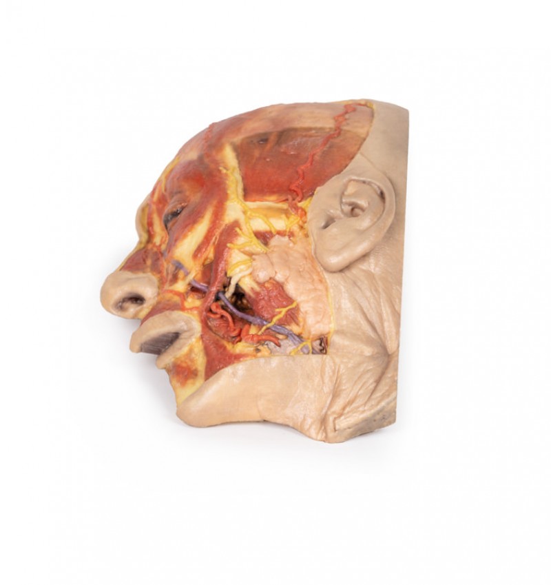
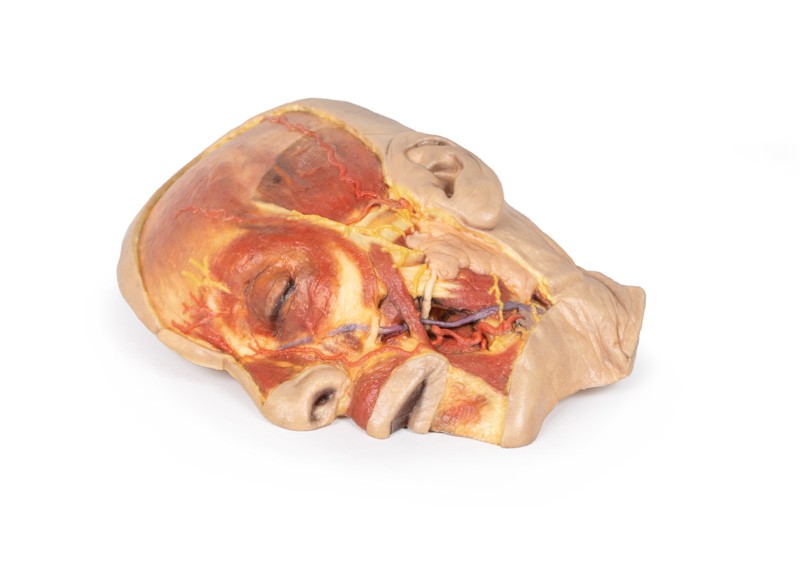
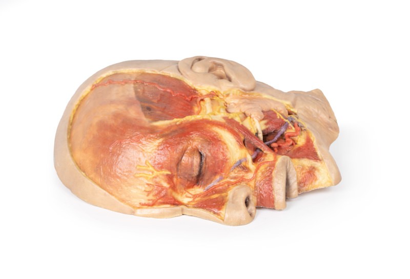
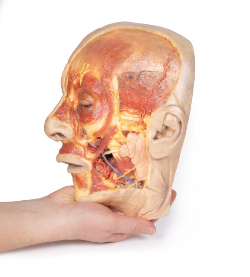
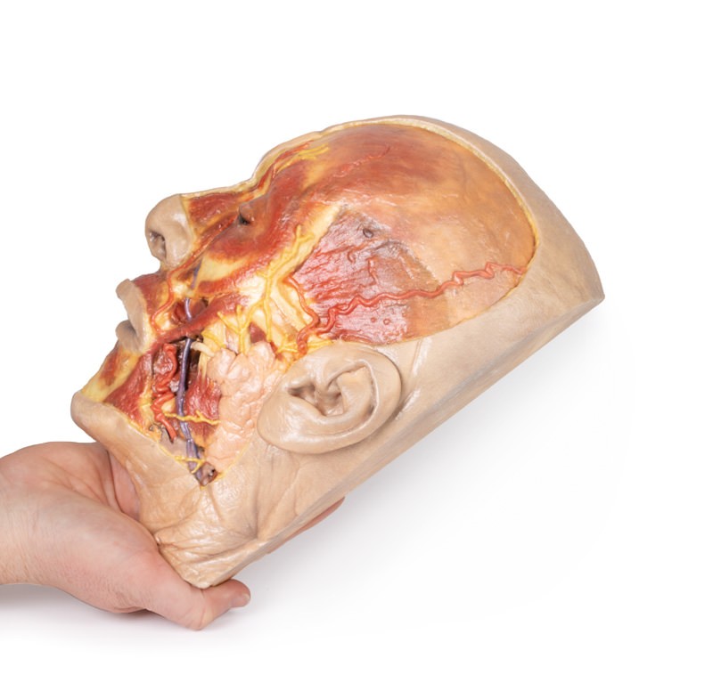
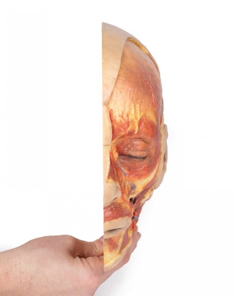
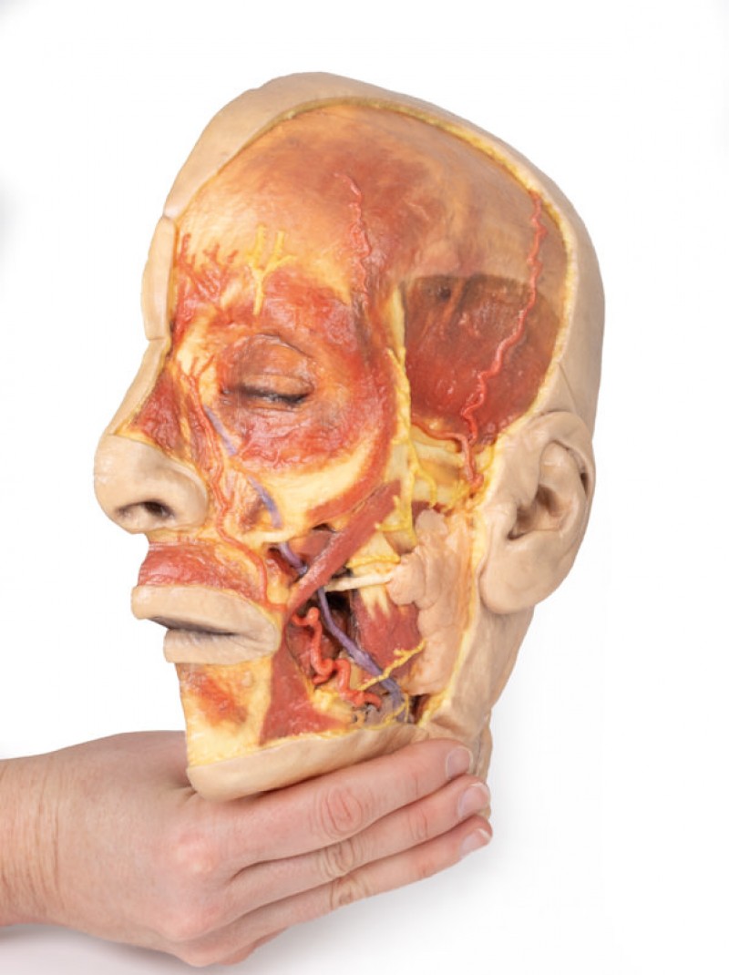
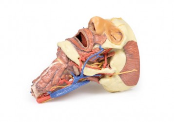
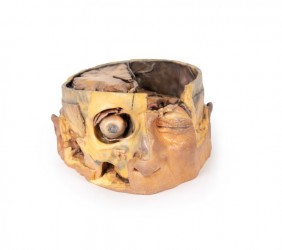
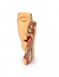
This 3D model presents a superficial dissection of a left face anterior to the ear with false colouring highlighting a series of neurovascular structures alongside the superficial muscles of facial expression. This compliments the more expanded superficial dissection of the face and lateral head presented in our HW 45 model. The undissected regions of the model have been digitally removed. Starting just anterior to the ear, the opened window of dissection has exposed the parotid gland and associated duct transmitting anterior towards the oral cavity. Exiting from the margins of the parotid gland are terminal branches of the facial nerve (CN VII), including the cervical, mandibular, buccal, zygomatic and temporal. The cervical and mandibular branches at the inferior portion of the dissection window can be seen angling inferiorly and passing superficially relative to the facial vein (which ascends towards the medial canthus of the eye). The mandibular branch passes just deep to the facial artery, which runs in parallel with the facial vein. Tracing the pathway of these vessels from the mandible towards the nasal and orbital regions also provides a checklist of superficial and deep muscles that have been highlighted, from the masseter deep to the parotid through to the depressor anguli oris, depressor labii inferioris, the zygomaticus major and minor, the orbicularis oris, the nasalis and levator labii superioris alaeque, the procerus and the orbicularis oculi.
Along the superior margin of the parotid gland the base of the auriculotemporal nerve and the superficial temporal artery ascends anterior to the ear and rests on a partially dissected temporal fascia to expose part of the temporalis muscle. Moving anteriorly over the orbit, the supraorbital nerve and supraorbital and supratrochlear arteries have been highlighted and ascend on the epicranial aponeurosis. Within that layer the deeper frontalis muscle can be appreciated as a darker shadow within the layer.
Inquiry
Related products
