


Home / 3D anatomy models / 3D head model / Parasagittal Section of the Head and Neck
Parasagittal Section of the Head and Neck
Parasagittal Section of the Head and Neck
 Download a PDF file Add to quotation - wish list
Product description: Parasagittal Section of the Head and Neck

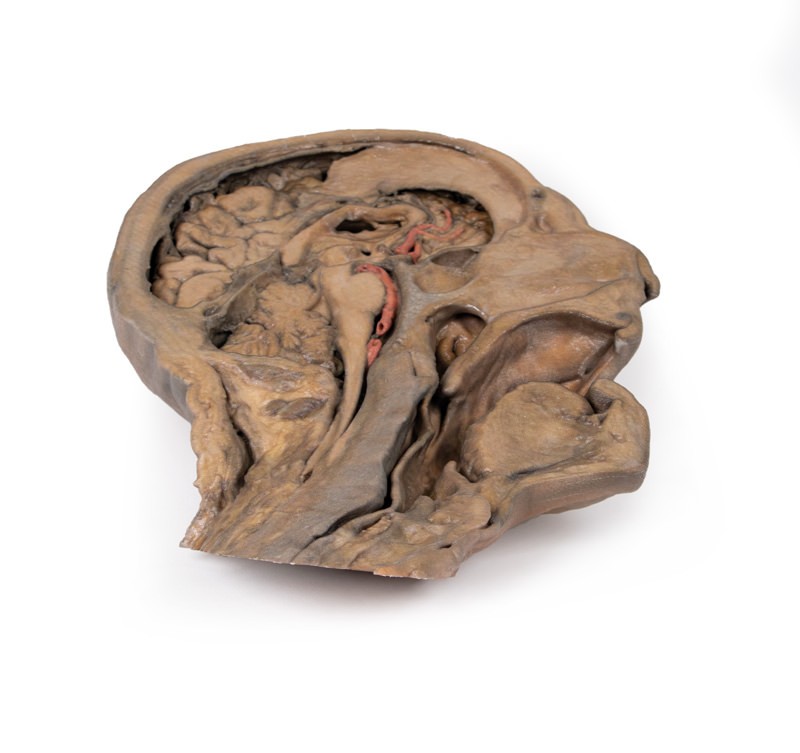
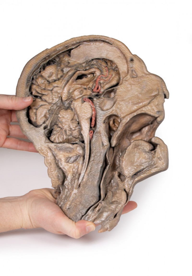
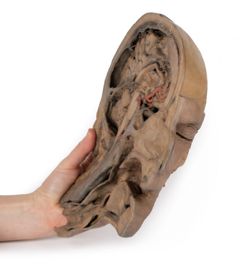
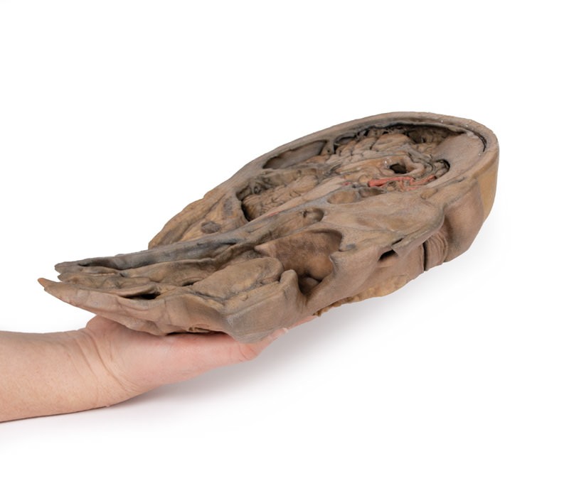
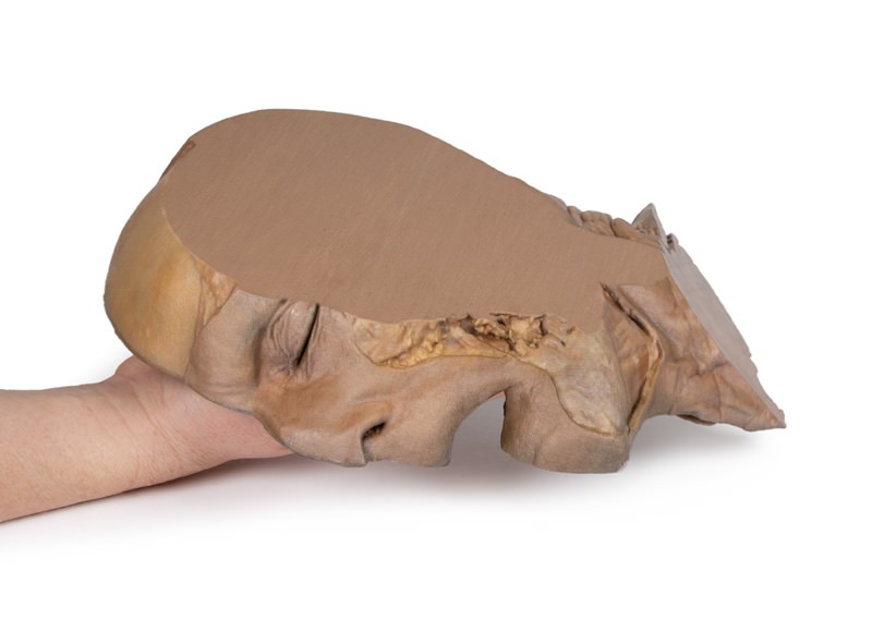
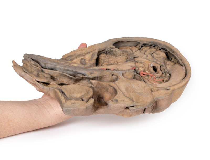
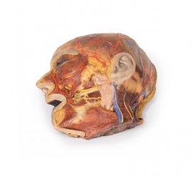
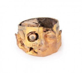
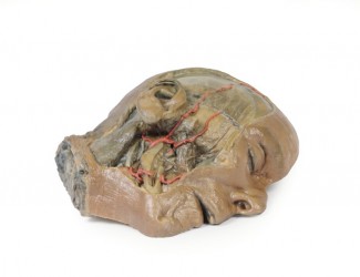
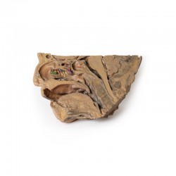
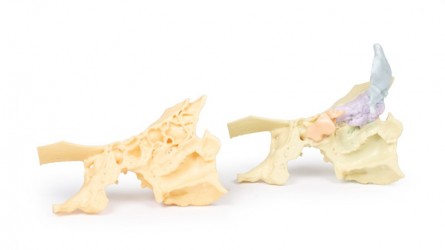
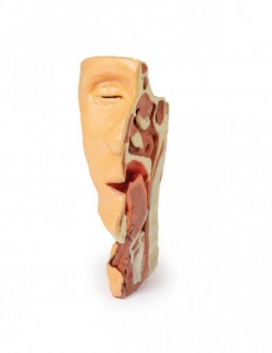
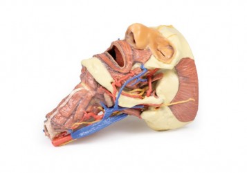
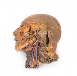
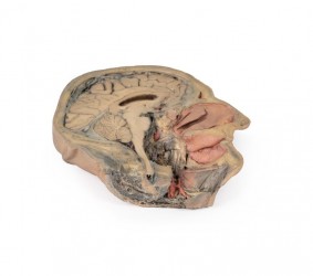
This 3D model of the head and neck represents a specimen sectioned just off the midsagittal plane to retain some midline anatomical structures (e.g., the falx cerebri, the septum pellucidum, the nasal septum) that are absent from other specimens in the series. There has also been fixative-induced shrinkage of the neural tissue. This reduction in volume has the benefit of exaggerating the space between the brain and endocranial contours and structures which are normally in closer approximation. The undissected side of the specimen has been digitally removed. The anterior part of the falx cerebri has been retained from its anterior attachment at the crista galli to roughly the midpoint of its extent towards the tentorium cerebelli. At the attachment of the falx part of the dura has been removed to demonstrate the extent of the superior sagittal sinus within the retained portion of the dural infold. The brain itself has been sectioned with preservation of the septum pellucidum and the interventricular foramen (of Monro) defining the passageway between the deep lateral ventricle and sectioned third ventricle. This section plane also captures the infundibulum extending from the hypothalamus to the pituitary gland, which is seated adjacent to a well-developed sphenoid sinus. Both the cerebral aqueduct and fourth ventricle are preserved, as are parts of the left vertebral artery, left posterior cerebral (in crosssection) and the branches of the anterior cerebral artery passing around the corpus callosum. The retention of the nasal septum in this specimen (and in contrast to other 3D models of the head and neck in the series) allows for an appreciation of the relationship between the septum and the hard and soft palates, the entrance of the auditory tube, and the overall nasopharynx relative to the nasal cavity and oropharyngeal region inferior to it. The muscular wall of the pharynx has been isolated to demonstrate the position relative to the cervical vertebral column. Inferiorly, the tracheal cartilages including the epiglottis, arytenoid and thyroid have been retained to demonstrate the position of these cartilages relative to the hyoid bone, as well as the vestibule, vestibular fold, and vocal fold in cross-section.
Inquiry
Related products