


Home / 3D anatomy models / 3D head model / Sinus Pathways
Sinus Pathways

Sinus Pathways
 Download a PDF file Add to quotation - wish list
Product description: Sinus Pathways

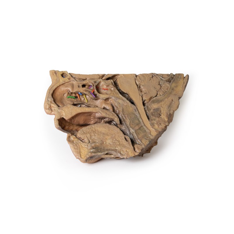
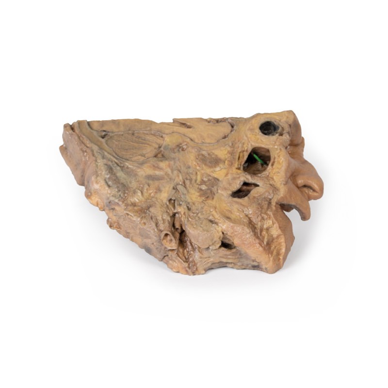
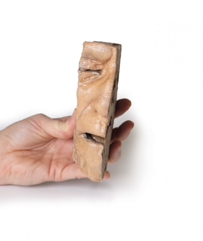
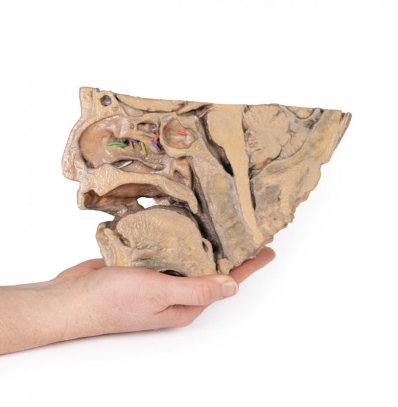
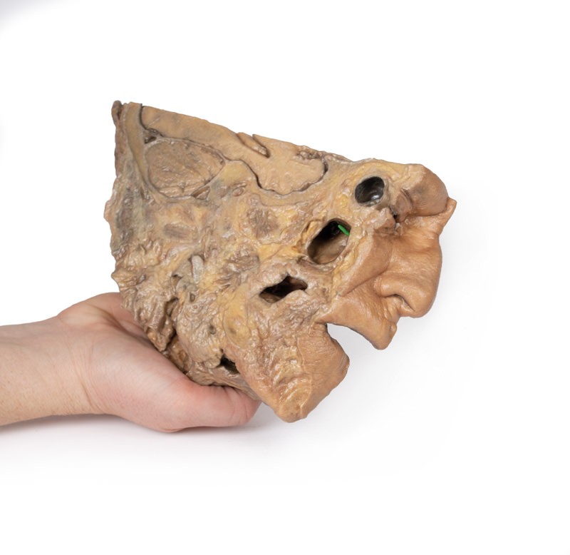
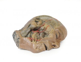
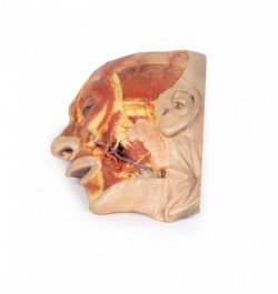
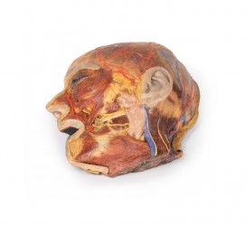
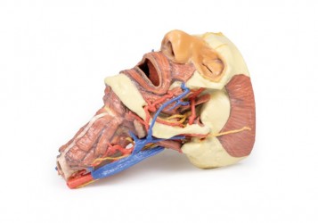
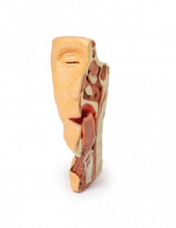
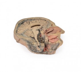
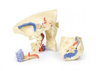
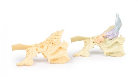
This 3D model provides a midsagittal to parasagittal segment of a right head to demonstrate the relationships and passageways of the paranasal sinuses. These passageways have been highlighted with thin coloured markers to indicate the relationship of these communicating routes between the paranasal sinuses and the nasal cavity. Starting anteriorly in the nasal cavity, the opening of the nasolacrimal duct (white) is present just deep to the inferior nasal conchae. The middle nasal concha has been sectioned to allow for a clear view of the opening of the maxillary sinus (visible in the parasagittal plane) across the semilunar hiatus (green), as well as the drainage of the frontal sinus (blue; with the sinus visible superiorly in the section and in the transverse cut through the specimen) and the anterior (orange) and middle (yellow) ethmoidal cells. The opening of the posterior ethmoidal cells into the superior meatus is shown through the purple marker, which is visible within a small opened window into the ethmoid just superior to the nasal cavity. Finally, the opening of the sphenoid sinus is marked in red and visible through the opened sphenoid sinus itself just superior to the nasopharyngeal region. In addition to these pathways, this 3D model also captures some of the surrounding anatomy within the section. Visible in the midsagittal view are the other primary structures of the nasal cavity from the nostril to the opening of the auditory tube posteriorly. The soft palate and uvula are preserved, as is the rest of the pharynx just to the level of the epiglottis and collapsed laryngeal region at the inferior part of the preserved specimen.
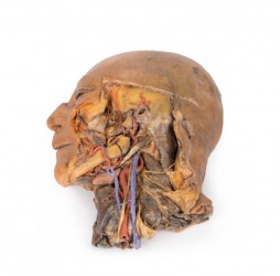
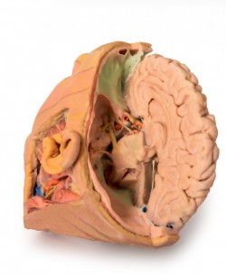
The oral cavity is displayed in cross section, with distinct genioglossus and geniohyoid muscles. In the cranial cavity, parts of the brain are preserved including the inferior parts of the frontal lobe and the right optic nerve/ chiasm/tract. The pituitary gland is visible in cross-section just superior to the sphenoid sinus. The pons, medulla oblongata, and most of the cerebellum are present, with a small part of the tentorium cerebelli separating the cerebellum from the right occipital lobe of the cerebrum. On the parasagittal side of the specimen there is a continuation of the tentorium cerebelli separating these parts of the brain, with clear cross-sections of the transverse sinus and part of the sigmoid sinus on either side of the cerebellum. Overlying this is a small part of the medial temporal lobe of the cerebellum with part of the anterior horn of the lateral ventricle deep within the lobe.
Inquiry
Related products