


Home / Anatomical Models / Anatomical Skull Models / Osteopathic Skull Model, 22 part, didactical version
Osteopathic Skull Model, 22 part, didactical version


Osteopathic Skull Model, 22 part, didactical version
 Download a PDF file Add to quotation - wish list
Product description: Osteopathic Skull Model, 22 part, didactical version

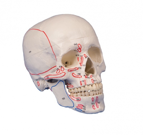
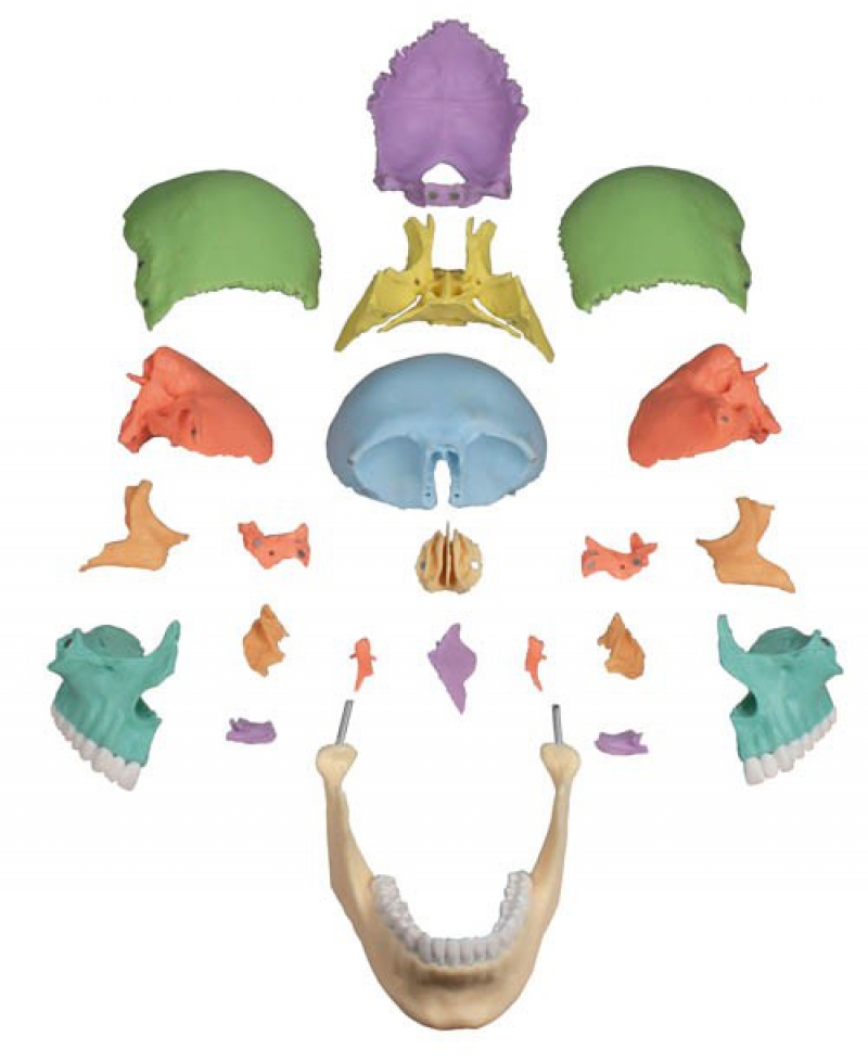
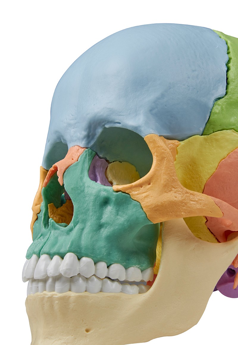
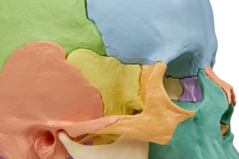
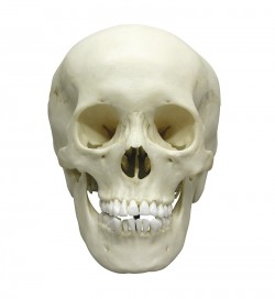
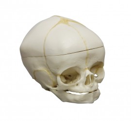
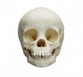
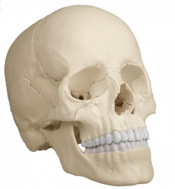
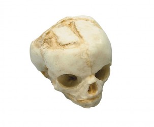
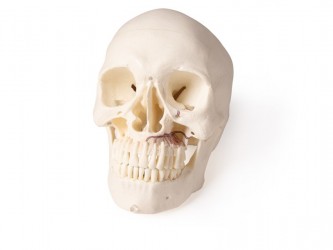
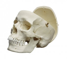
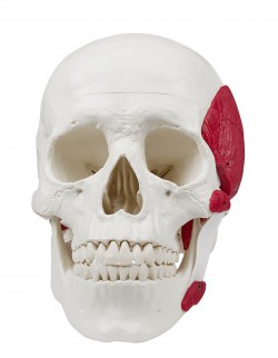
This fascinating natomical model of an average European adult skull can be disassembled into 22 single bones. During development of this model one of the main targets was to make the model easy to assemble and dismantle. Stable parts with convenient magnet connections make handling of the product a child‘s play. The detailed bones do not need any complicated pins to be stuck into holes, they almost slide into position, guided by realistic bone sutures and held by strong magnets. The perfect tool for Osteopaths.
The following bones are represented:
- Parietal bone left and right
- Occipital bone
- Temporal bone left and right
- Sphenoid bone
- Frontal bone
- Ethmoid bone
- Vomer
- Palatine bone, left and right
- Inferior nasal concha left and right
- Maxilla with teeth, left and right
- Lacrimal bone left and right
- Nasal bone left and right
- Zygomatic bone left and right
- Mandible with teeth
Supplied with users guide in English and German as well as a CD with Key card document in Latin, German, English, French, Spanish, Portuguese, Italian, Polish, Russian, Arabic, Korean and Japanese.
Inquiry
Related products


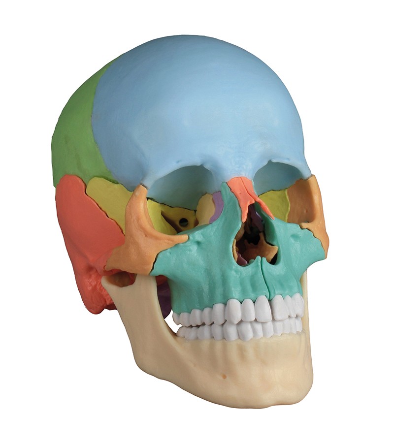
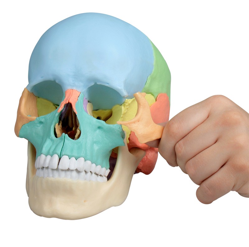
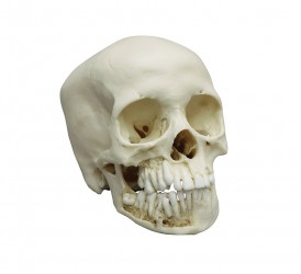
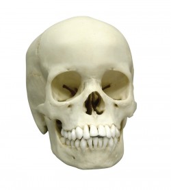
The anatomical model of the human skull, didactically colored, disassembled into 22 parts is an excellent teaching aid for everyone who wants to learn the details of the anatomical structure of the human skull. The use of different colors to paint individual elements makes it easier to learn the topography of the skull bones and the relationship between them. Thanks to the use of discreetly hidden magnets, you can easily disassemble and assemble the skull model, analyzing the structure of individual bones adequately to current needs.
The osteopathic skull is ideal for learning anatomy at home and during seminar classes at universities or medical schools. The anatomical model is manufactured by the German company Erler-Zimmer, the leader of this type of solutions on the global market.
Erler-Zimmer is a recognizable and world-renowned manufacturer of anatomical models, medical simulators and phantoms as well as veterinary models and simulators. For over 70 years it has been operating in the production and distribution of teaching aids in the field of all medical faculties and veterinary medicine. Erler-Zimmer anatomical models are made with the utmost care and from the best quality materials. Many of them are hand-finished, which is why Erler-Zimmer anatomical models are characterized by high accuracy and careful workmanship. Thanks to the use of modern 3D printing technology, Erler-Zimmer has created a unique series of 3D anatomical models. Based on radiological data, a series of models faithfully reflecting the human body was created. It is a modern solution used in many countries around the world.
Advantages of the osteopathic skull anatomical model:
- Can be disassembled into 22 separate parts
- Learning the anatomy of the normal bones of the skull
- Learning the topography of the bones of the skull and the relationship between them
- The individual elements are didactically painted Hand-finished
- Didactic CD included in different languages
Application of the skull model
- Anatomy lab equipment
- Learning the anatomy of the human skull at home
- The anatomical model can be used to demonstrate the structure of the human skull - to patients in office conditions
- As a teaching aid during postgraduate training/ courses for doctors, physiotherapists, osteopaths and other medical professions.