Quality Certyficate
street: Kolejowa 2, 30-805 Cracow


Home / Anatomical Models / Eye models
Anatomical Models
3D anatomy models
Custom tools for patient education
Veterinary simulators
Anatomical Charts
Anatomical Table 3D
Medical simulators
Medical Equipment
Type:
 See our profile on Facebook
 Check our profile on Instagram
 Download a PDF file
Anatomical Models / Eye models
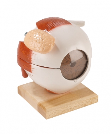
Featured on the exterior of the cornea are the large lacrimal gland, muscle attachments, optic nerve and blood vessels. To study interior features, the iris/cornea unit can be removed as can the functional lucite ...
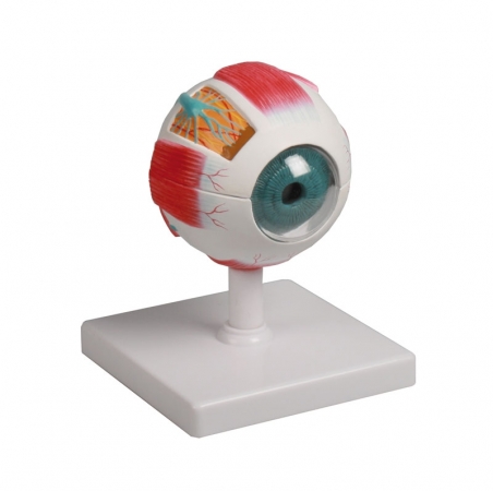
Model can be divided horizontally to show internal details. Cornea, iris, lens and vitreous body can be removed. Muscular attachments on the sclera and part of the choroid are also represented. Mounted on stand. ...
Model can be divided horizontally to show internal details. Cornea, iris, lens and vitreous body can be removed. Muscular attachments on the sclera and part of the choroid are also represented. Mounted on ...
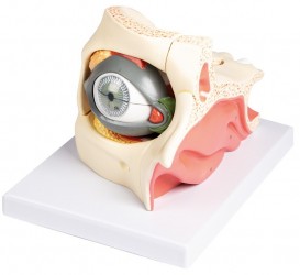
This 13-part model, enlarged approximately 2,5x life-size, shows the anatomy of the human eye.
The orbit can be opened into 3 parts to reveal the internal structures:
eyelid with lacrimal gland and duct
...
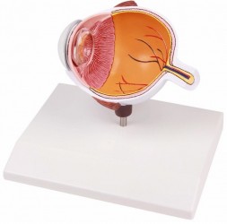
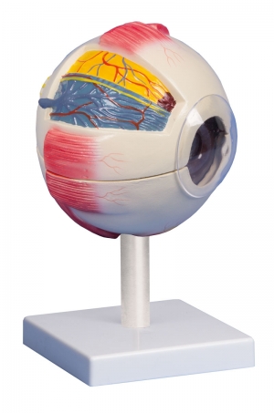
Cross section through the eyeball. The following anatomy is shown:
choroid
retina
macula
optic disc
optic nerve
central retinal artery and vein
retinal blood vessels
superior ...
1
Anatomical Models - Eye models
The organ of vision consists of the eyeball and additional organs. The eyeball is located in the eye socket, surrounded by the orbital fat body and has a spherical structure. The wall of the eyeball is made up of fibrous, vascular and retinal membranes. Inside the eyeball there is aqueous humor, lens and vitreous body. The accessory organs of the eyeball are the muscles and protective apparatus of the eyeball (eyebrows, eyelids, conjunctiva, lacrimal organ, fat body). The organ of vision is a complex anatomical structure, so anatomical models are perfect for educating about its structure and functions.
Anatomical models of the eye are useful teaching aids for everyone who wants to deepen their knowledge about the structure of the human eyeball. Using different colors to paint individual elements makes it easier to learn the topography of the anatomical structures of the human eye and the mutual relations between them. Thanks to the use of the highest quality materials and advanced production methods, human eye models are characterized by high durability and precision. Our assortment includes eye models made on a 1:1 scale and enlarged to better present individual structures. Some models can be broken down into parts, which makes the education process much more attractive and easier. Anatomical models constructed in this way are ideal equipment for anatomy laboratories.
Our offer includes, among others: the following anatomical models of the human eye:
- Eyeball model, 5 parts
- 6-piece eye model made with 4x magnification
- 6-piece eye model made with 6x magnification
- Cross-section (half) model of the eye
- Model of the eyeball and eye orbit
The human eye model is ideal for learning anatomy at home and during seminars conducted at universities and medical schools. Based on radiological data, a series of models faithfully reflecting the human body were created. These advanced teaching aids are the equipment of anatomy laboratories of many Medical Universities in Poland. This is a modern solution used in many countries around the world.
Advantages of anatomical models of the human eye:
- Accuracy of workmanship (hand-finished)
- Durability (made of the highest quality materials)
- Learning the normal anatomy of the human eye
- Learning the topography of anatomical structures located in the human eye and the mutual relations between them
- Eye models can be disassembled into parts, which makes the education process easier and more attractive
- Eye models made under magnification to better visualize anatomical structures
Application of human eye model:
- Equipment of the anatomy laboratory
- Learning the anatomy of the human eye
- The anatomical model can be used to demonstrate the structure of the human eye and eye pathology - to patients in an office setting
- As a teaching aid during postgraduate training for doctors, physiotherapists, osteopaths and other medical professions.
We also recommend skull models, ear models and tongue models, which, combined with anatomical eye models, will create an ideal set for learning anatomy.
If you have any questions or if the eye model you need is not available in the store, please contact us via e-mail or telephone. We are sure that together we will be able to provide the necessary materials for learning and improving skills. We can produce custom models with individual specifications.
