Quality Certyficate
street: Kolejowa 2, 30-805 Cracow


Home / Anatomical Models / Anatomical Skull Models
Anatomical Models
3D anatomy models
Custom tools for patient education
Veterinary simulators
Anatomical Charts
Anatomical Table 3D
Medical simulators
Medical Equipment
Type:
 See our profile on Facebook
 Check our profile on Instagram
 Download a PDF file
Anatomical Models / Anatomical Skull Models

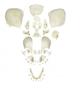
This fascinating natomical model of an average European adult skull can be disassembled into 22 single bones. During development of this model one of the main targets was to make the model easy to assemble and ...
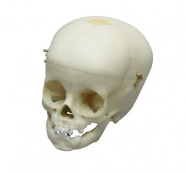
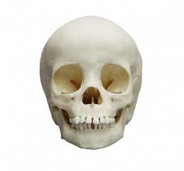
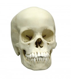
This skull model is a cast of a real human children‘s skull specimen and shows all anatomical structures in great detail. It was developed for students of anatomy, medicine, surgery, ENT medicine, ophthalmology and ...
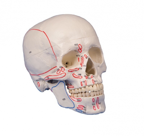
The human skull with marking of muscle insertions and origins. With nomenclature.
Size: 18 x 19 x 12 cm, weight: 0.7 kg ...
An excellent example of a condition in which the skull is abnormally long and narrow, as a result of premature closure of the sagittal suture, with heavy centers of ossification in the line of the suture.
Size: 20 x ...
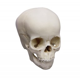
First class actual cast of a 15 months old child in extraordinary high detail.
Due to the very special production technology even smallest details are reproduced and the model looks and feels almost ...
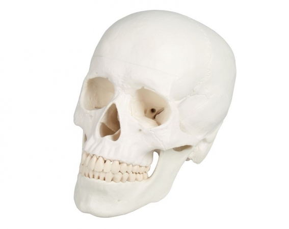
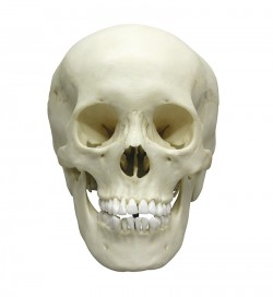
Developing this lifelike reproduction of a human skull we have used the latest technology to digitalize a real human skull and idealized it under the aspects of medical education. This means the skull was adjusted to ...
This fascinating model of an average European adult skull can be disassembled into 22 single bones.
During development of this model one of the main targets was to make the model easy to assemble and ...
A selected quality human skull was chosen for this model, dissectible into 14 parts: A horizontal section of the skull exposes the cranial cavity in which the course of the meningeal vessels, the venous ...
First class actual cast of a female human adult in extraordinary high detail. Due to the very special production technology even smallest details are reproduced and the model looks and feels almost like a real human ...
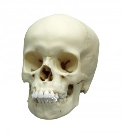
Skull model of a 5-year-old child. Extraordinary detail of anatomical details. Thanks to the special production technology, all major age-appropriate structures are reflected.
Additional information:
Size: 18 ...
First class actual cast of a female human 12 year old child in extraordinary high detail. Due to the very special production technology even smallest details are reproduced and the model looks and feels almost like a ...
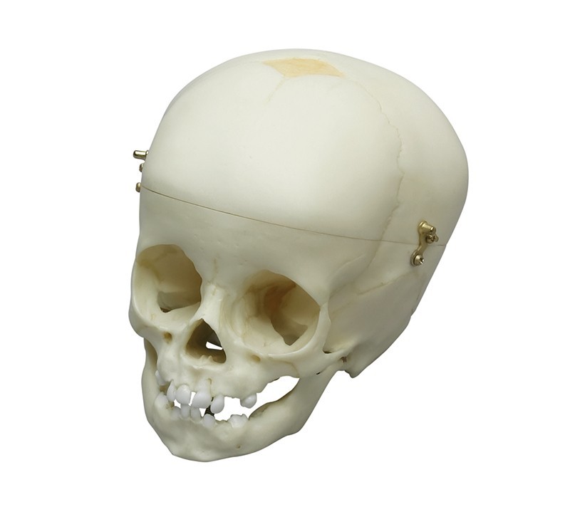
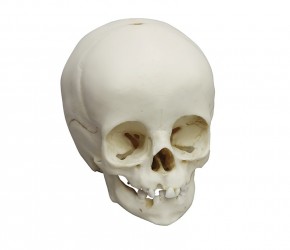
First class actual cast of a 1 year old child in extraordinary high detail and calvarian cut. Due to the very special production technology even smallest details are reproduced and the model looks and feels almost ...
First class actual cast of a male human adult in extraordinary high detail. Due to the very special production technology even smallest details are reproduced and the model looks and feels almost like a real human ...
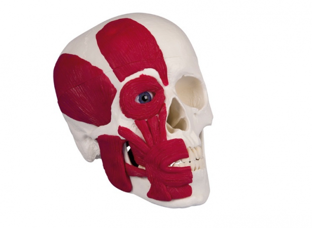
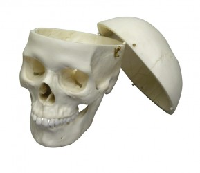
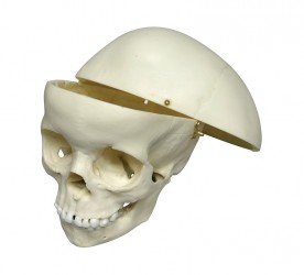
This fascinating model shows a human skull with the most important head muscles on one half. the mandible is unmovable. The skull cap can be opened and removed.
Size: 18 x 19 x 12 cm, weight: 0.8 kg ...
First class actual cast of a 3 year old child in extraordinary high detail.
This skull also shows numerous Wormian (sutural) bones found along the lambdoid suture.
The epipteric bone, which is ...
First class actual cast of a 4 year old child in extraordinary high detail. Due to the very special production technology even smallest details are reproduced and the model looks and feels almost like a real human ...
Skull model of a 9-year-old child. Thanks to the special production technology, the smallest anatomical details are reflected, thanks to which this model looks like a real skull.
Additional information:
Size: ...
First class actual cast of a 14 months old child in extraordinary high detail. Due to the very special production technology even smallest details are reproduced and the model looks and feels almost like a real human ...
Detailed skull model for learning anatomy designed for medical and dental students as well as other medical care. Thanks to the use of 3D printing technology and radiological data, this product contains numerous ...
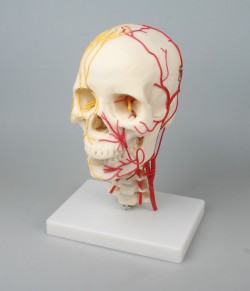
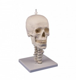
A life-size anatomical model of the human skull with vascularization. It presents the vascularization and innervation of the skull. Has 7 cervical vertebrae. On one side of the model arteries were presented, while on ...
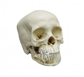
The teeth in the upper and lower jaw can be extracted and reinserted. The mandible is open and shows roots, spongiosa, nerve canal and an impacted wisdom tooth. The mandible is removable.
Size: 22 x 13 x 17 cm, ...
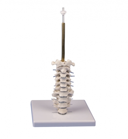
This cervical spine skull stand can be ordered as option for all skulls.
Since the skull has to be adjusted to the stand it may not be used to equip your existing skull.
Skull not included, ...
First class actual cast of a female human adolescent in extraordinary high detail. Due to the very special production technology even smallest details are reproduced and the model looks and feels almost like a real ...
This skull offers an excellent example of an adolescent. With the exception of the wisdom teeth, all permanent teeth are fully erupted, and no deciduous dentition remains. The apices of the canines, premolars, and ...
This disarticulated skull is from a full term (10 lunar months) fetus. It was part of a medical examiner‘s comparative pathology collection before going to the Maxwell Museum and is remarkable in its completeness. It ...

A very realistic model of a 38-week foetal skull manufactured from our unique bone-like material. The separate skull bones are clearly shown and meticulous moulding distinguishes such detail as sutures, fontanelles ...
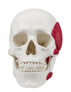
The masticatory muscles (masseter muscle, temporal muscle, pterygoideus medialis and lateralis muscles) are represented in the form of elastic bands. Using this model, it is possible to demonstrate the function of ...
Developing this lifelike reproduction of a human skull we have used the latest technology to digitalize a real human skull and idealized it under the aspects of medical education. This means the skull was adjusted to ...
12 partial anatomical model of human skull. High accuracy of performance, based on the analysis of radiological images, the structure of real skulls and literature.
The model is divided into: frontal bon, ...
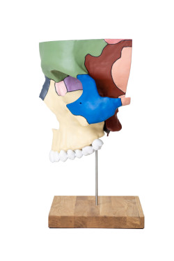
Didactic anatomical model of the middle of the left jaw made with a 4x magnification. The bones of the left craniofacial side are shown in different colors to facilitate the education process. The model has the ...
First class actual cast of a 1 ½ year old child in extraordinary high detail. Due to the very special production technology even smallest details are reproduced and the model looks and feels almost like a real human ...
Perfect reproduction of the fetal skull. At first glance, this model is hard to distinguish from the real skull and gives an impressive overview of the development of the skull in the womb.
Specification:
...
Anatomical Models - Anatomical Skull Models
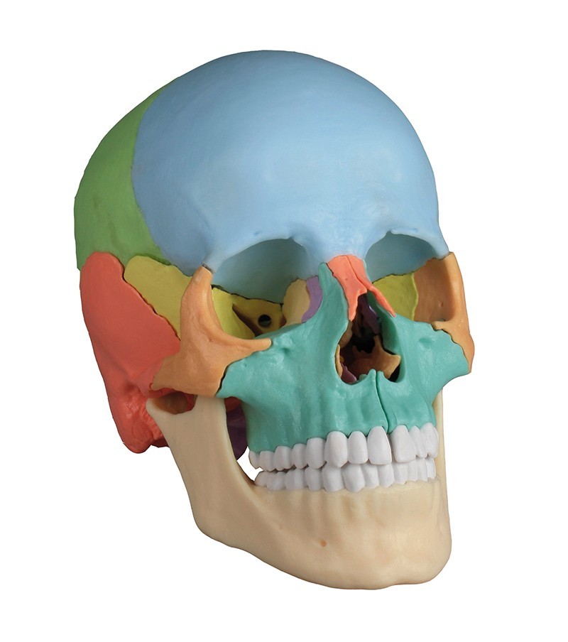
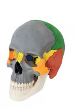
The human skull model is a teaching aid presenting the anatomical structure of the human skull. Our product range includes various anatomical models of the human skull. Depending on the level of advancement, we distinguish 1-piece and multi-piece skull models. The most advanced skull model in our offer in terms of the number of elements is made of 22 elements connected together using discreetly hidden magnets. It is an osteopathic skull. It is available in two variants - colorful (didactic version) and white (anatomical version).
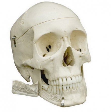
Due to the type of production technique, it is possible to obtain a more or less detailed model of a human skull. Some of our products are real casts of human skulls. They have very specialized anatomical details required when learning anatomy or planning a procedure. An example of such a product is a partially assembled human skull for medical research. This top-notch anatomical skull consists of 22 separate cranial bones and 32 individual teeth. The skull model is assembled bone by bone from a completely unassembled version and is held on a stand along with two separate parietal lobes to allow access to the interior of the skull. 32 teeth can be removed individually. This is a model of a young woman's skull, it is very detailed and shows all the holes, channels, seams and the smallest details of a real skull.
Other skull models in our assortment are created based on the use of radiological data from CT and modern 3D printing technology. This type of educational aid is, for example, an anatomical skull model made of 4 parts. This is a detailed skull model for learning anatomy, intended for students of medicine, dentistry and other medical fields. Thanks to the use of 3D printing technology and radiological data, this product contains numerous anatomical details necessary for learning anatomy in medicine. The model consists of a total of 4 parts that are connected together using magnets embedded in the material. The described skull model is part of the equipment of the anatomy laboratories of Medical Universities with a medical faculty and is recommended by anatomists conducting classes at Medical Universities. Our offer also includes another 4-piece skull model, dedicated to dentists. The dental skull has the ability to dismantle individual teeth, a movable and opening jaw with the possibility of presenting the nerve canal. Another demonstrative model of a dental skull should also be mentioned here. This is a 14-piece model of an adult human skull. The horizontal section shows the skull cavity, inside which the courses of the meningeal vessels, the venous sinus and the internal carotid artery are marked in color. In the sagittal section, the model shows the nasal cavity, frontal sinus and nasal sinus. It has the ability to dismantle the temporal bone and present the inner ear. The described skull model has a movable mandible with the possibility of disassembly and presentation of tooth roots and the nerve canal. It is worth noting that there is also a luxurious model of a human skull. It consists of 14 elements and is intended for advanced learning due to its numerous details and method of execution. The model is a cast of a real human skull and shows all anatomical structures in detail. It was developed for teaching anatomy for students of medicine, surgery, ENT medicine, ophthalmology and dentistry. The skull is carefully crafted and connected with metal and magnetic contact elements. The skull vault was divided in a horizontal plane, leaving the temporal bone and its sutures intact. Color-coded bony impressions of the superior sagittal sinus, transverse and sigmoid sinuses, and meningeal vessels. The base of the skull model has been divided in the sagittal plane so that it passes through one ethmoidal plate on one side, and another section in the same plane passes through the other ethmoidal plate, leaving intact the perpendicular ethmoid plate crista galli as well as the entire nasal septum. The anterior, middle, and posterior structures of the cranial fossa are easily accessible. The nasal cavity, turbinate, nasal septum, bony pharyngeal space and nasopharynx can be directly presented. The oval window, semicircular canals and the opening of the tympanic sinus are visible. In the maxilla and mandible, dental structures, roots, the bony edge of the alveolar process, dental vessels and nerves are visible. The maxillary sinus can be opened by removing a bone flap. The teeth of the right lower jaw are cut to show the internal tooth structure.
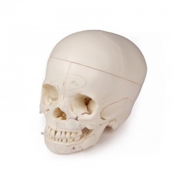
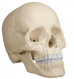
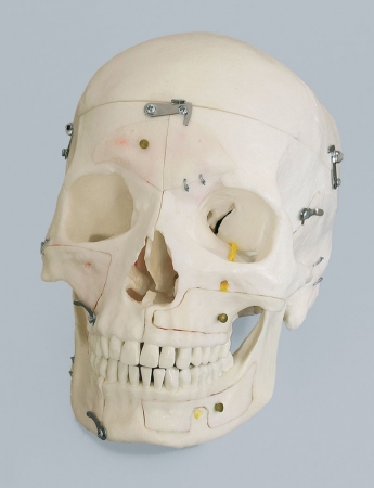
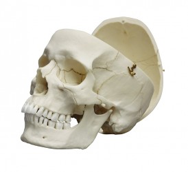
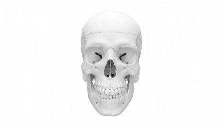
As a supplier wishing to meet the expectations of all customers, we also offer models of the human skull with a less complicated structure. As an example, there is a skull model popular on the Polish market, consisting of 3 parts. In developing this realistic reproduction of a human skull, the latest digitization technology was used to create a model specifically intended for medical education. The described model can be disassembled into 3 parts: cover, base and jaw. The teeth correspond to the actual, natural dentition regarding their position. Thanks to the movable and removable jaw, it is possible to demonstrate mobility in the temporomandibular joints. The skull cover fits securely to the base with metal pins and strong, discreetly placed magnets. Thanks to this solution, impractical and unsightly protruding pins and hooks that were previously used in this type of teaching aids were dispensed with. This is a feature that distinguishes this human skull model from other similar educational tools available on the market. Another, more advanced 3-piece model of the human skull is a version with additional hand-made markings of the skull muscle attachments. It is delivered with an educational card in English and German. Our assortment also includes a version of the skull model with numbered bone elements, which undoubtedly facilitates learning the topography of the skull bone structures and the mutual relations between them. It is worth mentioning the skull model with facial muscles. This is a life-size bony anatomical model of an adult human skull with the major facial muscles depicted on one side. The model has a removable cover and limited mobility in the temporomandibular joints.
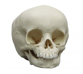
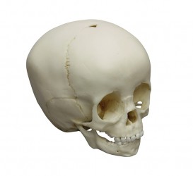
Anatomical model of a child's skull, a model of an infant's skull, a model of a newborn's skull:
It should be noted that the skull of infants looks completely different than that of an adult. In newborns, the skull bones still contain soft, unossified elements. These are the remains of a membranous skull. They call them fontanelles. Due to significant differences in the anatomical structure of infant skulls, it was decided to create models of children's skulls with features characteristic of a given period of life. They allow us to better understand the anatomical structure of the child's skull and trace its development from the moment of birth. The child's skull models have characteristic elements such as the frontal, occipital, sphenoid and mastoid fontanel, as well as other typical details appropriate to the child's age, such as erupting teeth, milk teeth, and dentition characteristic of a given period of life. Our offer includes various types of models of a child's skull, from 1 to 15 years old. For example, a male human skull model, 13 years old, is a detailed anatomical aid. Thanks to special production technology, the smallest details are shown, which is why this model resembles a real skull of a 13-year-old child. With the exception of wisdom teeth, all permanent teeth are fully formed and no primary teeth remain. The tips of the canines, premolars, and second molars are approximately one-half to two-thirds radiographically closed; this is consistent with the age of 13.5 - 14 years. The crowns of the three remaining third molars are in the early stages of calcification. Another model of a children's skull is the skull of a 5-year-old child. It is characterized by extraordinary detail of anatomical details. Thanks to special production and dyeing technology, all the most important age-appropriate structures were reflected. The skull model has the following dimensions: 18 x 12 x 13 cm.
Fetal skull model:
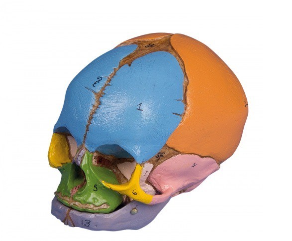
The collection of realistic fetal skulls is undoubtedly worth attention. Our offer includes models of fetal skulls from the 13th week of pregnancy to the 40th week of pregnancy. The Didactic 38-Week Fetal Skull is one of the most popular models in this collection. The described product is an extremely realistic model of the skull of a 38-week-old fetus made of bone-like material. The individual elements of the skull are presented in detail. The model shows the skull sutures, fontanelle, and external auditory canal. The main bones are presented in different colors for easier identification. The model comes with an educational card in English.
The osteopathic skull and other anatomical skull models from our offer are ideal for learning the anatomy and topography of the bone structures of the skull. It is an excellent educational tool for medical students, physiotherapists, osteopaths and doctors. This type of skull model is also a valuable and multifunctional equipment for specialist offices or medical facilities because it can be used to help explain to patients the essence of the problem and how to treat it.
Adult skull anatomy:
The human skull is an important bone structure that protects the brain and the organs it contains. The bones of the skull are divided into the bones of the cerebral part and the facial bones. The skull also has an upper wall, a lower wall, a front wall, a rear wall, and two lateral walls. The upper wall - the vault of the skull - is formed by the frontal scale (bone), two parietal bones and the upper part of the occipital bone. The skull base (lower wall) has an inner and outer surface. The outer surface of the skull base is formed, among others, by: through the bony palate, the lower surfaces of the greater wings of the sphenoid bone, the lower surfaces of the shaft of the sphenoid bone, the lower surfaces of the pyramids of the temporal bones and the lower surfaces of the occipital bone. The occipital bone has a foramen magnum and 2 occipital condyles. The inner surface of the skull base is divided into three fossaes: anterior middle and posterior. The frontal wall of the skull (facial skull) consists of the frontal bone, nasal bones, zygomatic bones, maxilla and mandible. The respiratory system and the digestive system originate in the anterior wall, as well as the sensory organs (smell, taste, sight). The organ of vision is located in the eye sockets, the organ of smell is in the nasal cavity and the organ of taste is in the oral cavity. The back wall of the skull is formed mainly by the occipital bone and partly by the parietal and temporal bones. The lateral wall of the skull is formed by the temporal bone, the zygomatic bone and the sphenoid bone. On the lateral wall there is a zygomatic arch and the external auditory foramen. The connections of the skull bones are diverse. These are sutures, cartilage, osteochondrosis and joints. The temporomandibular joint is a paired joint formed by the head of the mandible and the mandibular fossa of the temporal bone, it contains an articular disc that creates a movable socket for the joint. These joints perform the movements of chewing, opening and closing the mouth, and extending and retracting the jaw.
Osteopathic Skull:
The most popular skull model in our offer is the Osteopathic skull. This 22-piece skull model has educationally colored bones (each in a different color). This makes it easier for students to learn the anatomy and topography of the skull, as well as the interrelationships of the bones that make it up and the connections between them. This model allows you to easily assemble and disassemble individual elements, which are connected together in a practical way using discreetly hidden magnets. This is an ideal skull model for learning anatomy for doctors, physiotherapists and osteopaths. The skull model is supplied with instructions in English and German as well as a CD with documentation in Latin, German, English, French, Spanish, Portuguese, Italian, Polish, Russian, Arabic, Korean and Japanese.
The skull model described above contains the following bones:
- Parietal bone left and right
- Occipital protuberance
- Temporal bone right and left
- Sphenoid bone
- Frontal bone
- Ethmoid bone
- Blade
- Right and left palatine bone
- Right and left inferior turbinates
- Jaw with teeth
- Lacrimal bone right and left
- Nasal bone right and left
- Right and left zygomatic bone
- Mandible with teeth
Anatomical models of the skull - application:
- learning anatomy
- learning the topography of the bone structures of the skull
- anatomy laboratory equipment
- equipment for biological and natural science laboratories in schools, technical schools, universities, and medical offices
- teaching aid for medical students
- educational tool for patients in the office setting
