Quality Certyficate
street: Kolejowa 2, 30-805 Cracow


Home / Anatomical Models / Human Skeleton Models
Anatomical Models
3D anatomy models
Custom tools for patient education
Veterinary simulators
Anatomical Charts
Anatomical Table 3D
Medical simulators
Medical Equipment
Type:
 See our profile on Facebook
 Check our profile on Instagram
 Download a PDF file
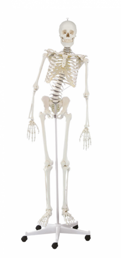
Human skeleton model - miscellaneous
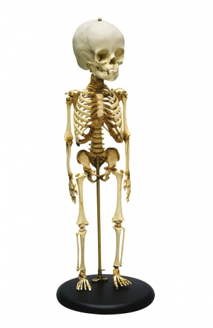
Child skeleton model
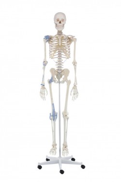
Adult skeleton model
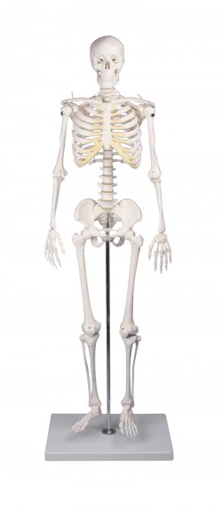
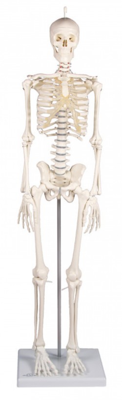
Miniature skeleton
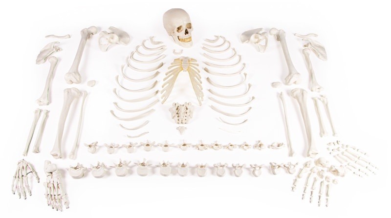
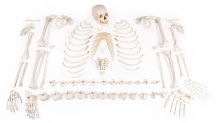
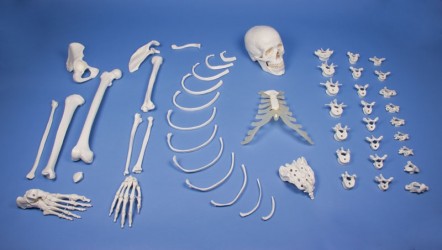
Skeleton disassembled
Anatomical Models / Human Skeleton Models
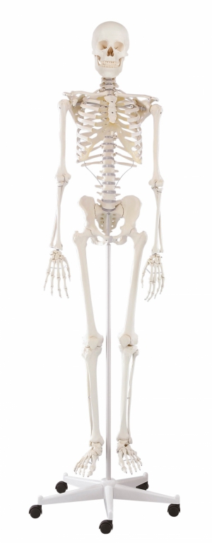
School skeleton. A full-size model of the human skeleton intended for educational purposes. It allows you to demonstrate the anatomical structure of the skeletal system, topography of individual bones and their mutual ...
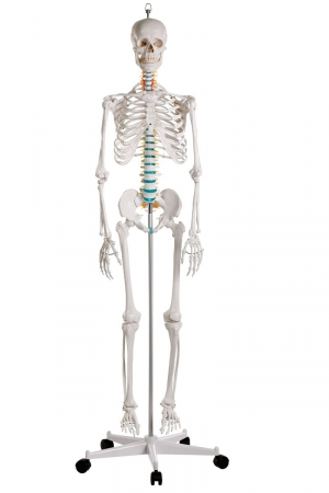
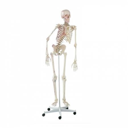
This therapy skeleton with a moveable vertebral column is ideal for anyone who not only wants to learn anatomy, but also wishes as a therapist to understand or explain the connections between movements, postures and ...
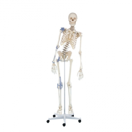
This therapy skeleton offers articular ligaments on one body side in addition to the moveable vertebral column.
The model has the following characteristics:
Natural casting of a human skeleton
...
Anatomical skeleton with muscle marking. All anatomical structures and details are represented in detail.
The model has the following characteristics:
Natural casting of a human skeleton
Representation of ...

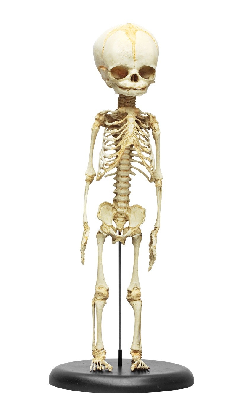
A detailed skeleton of a human fetus on the base.
Average body length measurements of this skeleton suggest an age of 8.5 to 9 months, but developmental (non-metric) osteological traits are most suggestive from 7 to ...
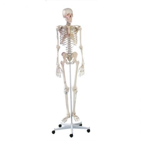
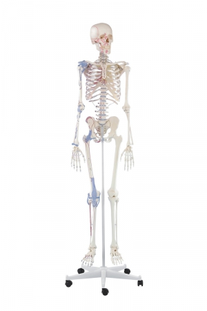
This therapy skeleton offers marking of the muscle origins and insertion points on one body side in addition to the moveable vertebral column.
The model has the following characteristics:
Natural casting of ...
Anatomical skeleton with articular ligaments as well as muscle marking. All anatomical structures and details are represented in detail.
The model has the following characteristics:
Natural casting of a human ...
Pristine examples of adolescent skeletons are rare finds in teaching collections. After much searching, we have discovered an excellent example of an adolescent skeleton. The developing skeleton is very different ...
Realistic skeleton of a child aged 5 years.
Characteristic:
The age of the skeleton is confirmed based on the pattern of eruption (eruption) of teeth and the developmental age of individual bones.
...
This therapy skeleton with a moveable vertebral column is ideal for anyone who not only wants to learn anatomy, but also wishes as a therapist to understand or explain the connections between movements, postures and ...
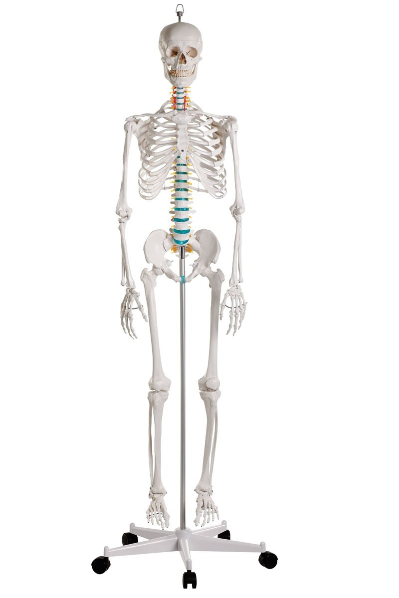
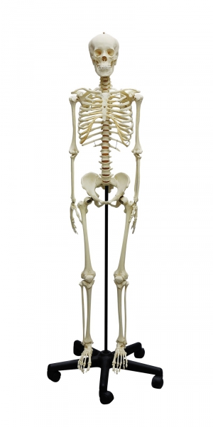
All bones in the skeleton are represented individually. Excellent casting of the skeleton of an adult male. True to life reproduction of the bone structure with all foramina, fissures and processes. The skull can be ...
Model of children's skeleton at the age of 14-16 months.
Characteristics:
It presents the epiphyses of the long bones and the cartilaginous periphery of many bones characteristic of this stage of ...
The model shows a twice-reduced human skeleton. Skulls, arms and legs are removed. In addition, the skull can be divided into three parts.
Specifications:
dimensions: approx. 84 cm high (without the base)
...
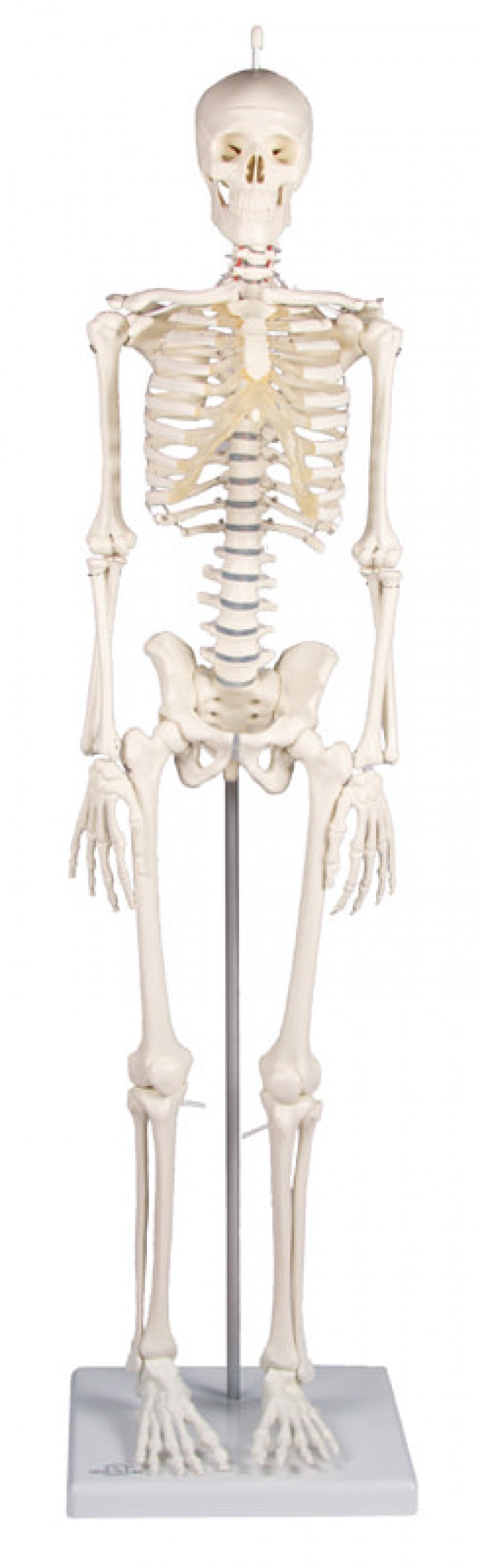
The model shows a twice-reduced human skeleton. The arms and legs are fully movable; shoulders, hips, knees and ankles are mounted as sliding joints to allow natural movement. The three-part skull and the arms and legs ...
The model shows a twice-reduced human skeleton. The arms and legs are fully movable, the arms, hips, knees and upper ankles are designed as sliding joints, so you can show all natural movements. The three-part skull, ...
The model shows a twice-reduced human skeleton. The arms and legs are fully movable, the arms, hips, knees and upper ankles are designed as sliding joints, so you can show all natural movements. The three-part skull, ...
The ideal model for anatomical study. All details and structures are faithfully reproduced.
The model has the following characteristics:
Natural casting of a human skeleton
Representation of all ...

...
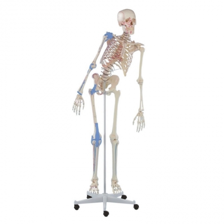
This therapy skeleton offers marking of the muscle origins and insertion points on one body side in addition to the moveable vertebral column and muscle marking.
The model has the following characteristics:
...
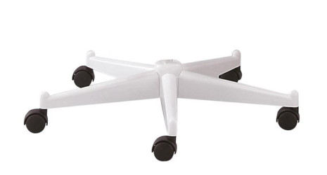
Tripod suitable for all life-size EZ skeletons, 5-beam WITHOUT tripod tube (outer diameter 18.0 mm). ...
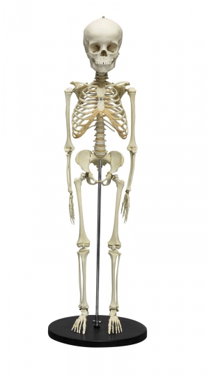
All bones on the right side or those occurring once in the skeleton are represented individually. Excellent casting of the skeleton of an adult male. True to life reproduction of the bone structure with all foramina, ...
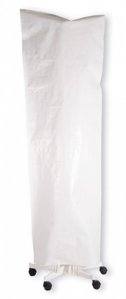
This dust cover is made of a durable plastic fabric, is tear-resistant and offers very good protection against dust build-up. The cover is simply pulled over the frame and is virtually indestructible as no moving parts ...
A 1/2 life-size scale model of the human skeleton. The arms and legs are fully movable; The arms, hips, knee and ankle are mounted as movable joints to allow for natural movement. The three-part skull as well as the ...
The model has the following features:
All anatomical details (e.g. holes, slots, joints)
A natural cast of the human skeleton
Skull disassembled into three parts
Removable ...
1
Anatomical Models - Human Skeleton Models
The human skeleton performs protective and supporting functions in the body, is a place for muscle attachment and is a passive part of the musculoskeletal system. An adult human has approximately 206 different bones. In order to facilitate learning the anatomical structure and biomechanics of the locomotor system, anatomical models of the human skeleton are helpful. These are teaching aids intended for everyone who wants to deepen their knowledge of osteology and joint mechanics. Using different colors to paint individual elements makes it easier to learn the topography of anatomical structures and the mutual relations between them. Thanks to the use of the highest quality materials and advanced production methods, human skeleton models are characterized by high durability and precision. Many anatomical models of the skeleton, e.g. presenting the bone skeleton, are real casts made of real human bones. Our assortment includes anatomical models of the adult skeleton and anatomical models of fetuses, children and teenagers. Depending on the type, anatomical models of the human skeleton present the skeleton as a whole or in fragments, e.g. a model of the chest. Hand-made markings of muscle attachments also bring great educational value. These types of anatomical models are delivered with an educational card containing the nomenclature of anatomical structures. Our assortment also includes anatomical models of skeletons made on a scale of 1:1 or in reduced size (desktop models made on a scale of e.g. 1:2).
Our offer includes, among others: the following anatomical models of the human skeleton:
- OSCAR - anatomical skeleton - basic version
- A human skeleton model for a physiotherapist!
- Toni's skeleton - with a movable spine and ligaments
- Arnold human skeleton with muscles marked
- Skeletal model of a 30-week fetus
- Peter human skeleton with movable spine and muscle markings
- Human skeleton model with marked muscles and ligaments - BERT
- Skeleton of a growing young man
- Skeletal model of a 5-year-old child
- Hugo - educational skeleton with a movable (flexible) spine
- Disassembled human skeleton
- Skeleton model of a child aged 14-16 months
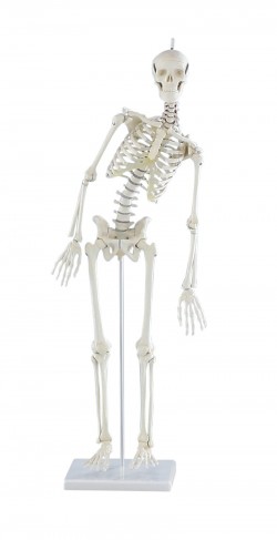
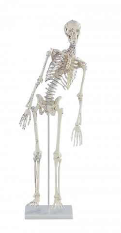
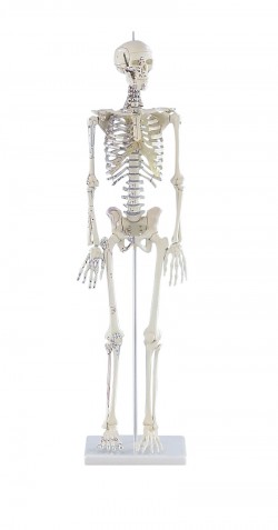
- Miniature human skeleton "Tom", 1/2 life size
- Miniature skeleton "Paul" with a movable spine
- Posable miniature skeleton "Fred" with muscle markings and a flexible spine
- Skeleton MAX
- "Otto" skeleton model with ligaments
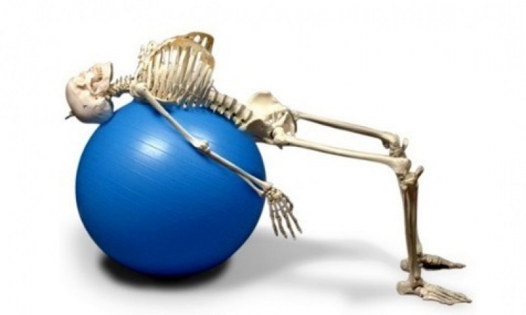
The human skeleton model is ideal for learning anatomy at home and during seminars conducted at universities and medical schools. The limbs are connected with elastic rubber, which allows for the presentation of anatomical and translational mobility in the joints. Some skeleton models have a flexible spine, which allows, for example, demonstration of chest movements, analysis of posture defects or demonstration of ball exercises. Flexible intervertebral discs in this type of models behave like natural ones. Virtually all models have removable limbs and skulls. The movable tripod included in the set makes it easier to move the model. Some models have hand-painted muscle attachments and flexible joint ligaments, which improves the teaching value of these precisely made educational aids. These advanced teaching aids are the equipment of anatomy laboratories of many Medical Universities in Poland. This is a modern solution used in many countries around the world.
Advantages of anatomical models of the human skeleton:
- Accuracy of workmanship (hand-finished)
- Durability (made of the highest quality materials)
- Learning proper anatomy
- Learning the topography of anatomical structures and the mutual relations between them
- Learning biomechanics of the musculoskeletal system
- Possibility to demonstrate posture defects
- Possibility to demonstrate exercises
- Learning muscle attachments
- Flexible intervertebral discs ensuring mobility of the spine
- A tripod on wheels for movement
- Some models have joint ligaments and marked muscle attachments
Application of human skeleton model:
- Equipment of the anatomy laboratory
- The study of skeletal anatomy, osteology
- The anatomical model can be used to demonstrate structure and injury/pathology to patients in an office setting
- As a teaching aid during postgraduate training for doctors, physiotherapists, osteopaths and other medical professions.
The anatomical models of the human skeleton that are in our offer are made of the highest quality materials. Thanks to their realistic structure, they enable quick and effective learning of anatomical structures and their functions.
We also recommend models of the pelvis and skull, which, combined with anatomical models of the human skeleton, will create an ideal set for learning anatomy.
If you have any questions or if the human skeleton model you need is not available in the store, please contact us via e-mail or telephone. We are sure that together we will be able to provide the necessary materials for learning and improving skills. We can produce custom models with individual specifications.
