Quality Certyficate
street: Kolejowa 2, 30-805 Cracow


Home
3D anatomy models
Anatomical Models
Custom tools for patient education
Veterinary simulators
Anatomical Charts
Anatomical Table 3D
Medical simulators
Medical Equipment
Type:
 See our profile on Facebook
 Check our profile on Instagram
 Download a PDF file
This 3D printed specimen compliments our dorsal dissection specimen (AM01273) by presenting a ventral deep dissection of axial anatomy from the head, neck, axillae, thorax, and abdomen to the proximal portion of the ...
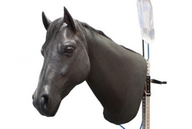
This advanced and realistic head simulator of equine on a tripod, designed for training and improving the skills of venipuncture and intramuscular injections.
Main features:
Jugular venipuncture with palpable ...
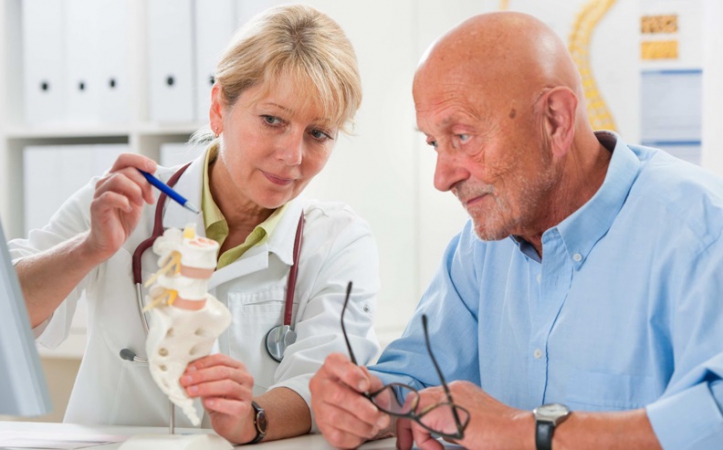
Patient's education increases the effectiveness of therapy!
Patients wants to know more about the disease they are suffering from. Also they want to know the way of therapy. Help your customer to improve the patient ...
This therapy skeleton with a moveable vertebral column is ideal for anyone who not only wants to learn anatomy, but also wishes as a therapist to understand or explain the connections between movements, postures and ...
Medical Device Replica
We offer a cost-effective alternative to your original device. The replicated devices can be used for marketing purposes or patient education Service.
Services
Custom anatomical ...
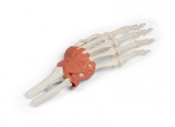
This model shows the wrist and carpal tunnel.
The following ligaments are shown, among others:
Retinaculum flexorum, Lig. pisometacarpum, Lig. pisohamatum, Tendo m. flexoris carpi radialis, Lig. ulnocarpale palmare, ...
The Facial simulator contains the following muscles:
Frontalis, Temporalis, O/Oculi, Risorius, Zygomaticus major & minor, Levator Labii, Buccinator, Alaeque Nasi, Orbicularis Oris, Labii Inferioris, Mentalis, ...
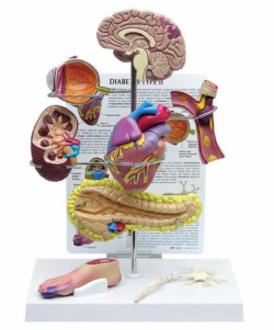
The includes miniature brain, eye, heart, kidney, artery, pancreas, neuron, and foot models. Education card illustrates effects associated with Type II Diabetes: stroke, ocular pathology, hypertensive heart disease, ...

The model simulates the correct and incorrect way of lifting weights for the spine. Ideally suited for demonstration purposes in cabinet conditions.
Size: 23 x 15 x 15 cm ...
Four piece model indicating structures and organs with vascular effects due to diabetes.
Includes sectioned model of Bowman‘s capsule (kidney), artery, nerve, eye (posterior section).
Full model size without ...
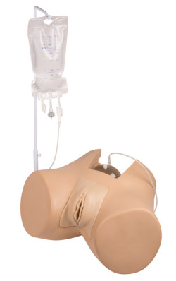
A professional training simulator for learning catheterization. This model is perfect for patient education.
Main features:
The innovative bladder is designed to provide a normal, anatomical and ...
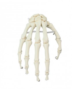
A life-size anatomical model showing the skeletal structure of the hand. It Shows all palm bones which are individually mobile-mounted on wire
It is possible to perform translational movements in individual hand ...
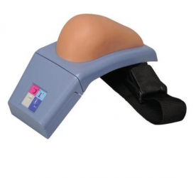
This high-quality I.M. simulator represents a right upper arm with all important anatomical palpable landmarks such as acromion and humerus. The realistic anatomy allows for placing correct intramuscular ...
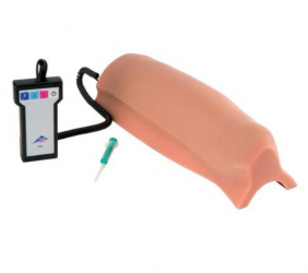
This high-quality simulation of a right upper leg realistically displays all of the important anatomical, palpable landmarks such as patella and greater trochanter for very realistic practice of correct ...
This high-quality, strap-on intramuscular simulator is a lifelike model of a right buttock with all important anatomical landmarks for intramuscular (I.M.) injections: iliac crest, anterior superior iliac spine and ...
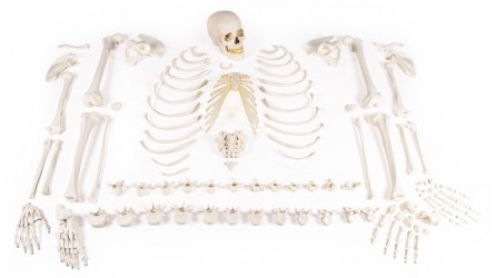
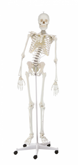
All bones in the skeleton are represented individually. Excellent casting of the skeleton of an adult male. True to life reproduction of the bone structure with all foramina, fissures and processes. The skull can be ...
The calving simulator is a practical tool for training a cow's delivery reception and carrying out ultrasound examinations. This compact trainer can be placed on a table at the correct height so that pupils / students ...
Our include 13 life-like magnetic models of pork cuts to help students learn to identify, locate, and prepare each piece. This pork carcass model, which is about half the size of a real pig, features realistic ...
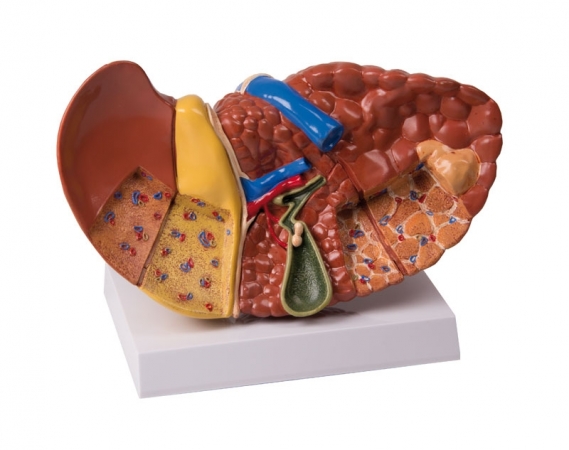
Full size liver model shows conditions such as: Cirrhosis (septal and nodular), biliary obstruction, gall stones, and tumors.
Size: 20 x 11.5 x 14 cm ...

The Stomach Model with Ulcers is a full size cut-away section of stomach shows gastric ulcer, duodenal ulcer and esophageal inflammation.
Model size: 7.75 x 2.25 x 6.25"
Base: 6.5 x 5" ...

1-Layer Suture Pad is designed for students to master knot tying and suture pattern uniformity. The transparent 3-dimensional view of the pad allows students to see and more fully understand the depth and accuracy of ...
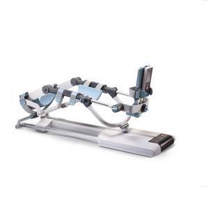
BTL-CPMOTION K ELITE
Ultimate all-in-one solution – single unit for all 3 leg joints
Remote control with touch screen
Advanced settings Main features
Ankle module
Precise real-time ...
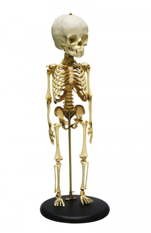
Model of children's skeleton at the age of 14-16 months.
Characteristics:
It presents the epiphyses of the long bones and the cartilaginous periphery of many bones characteristic of this stage of ...
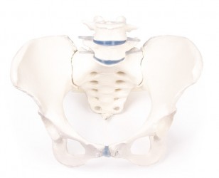
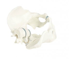
Natural casting of an adult female pelvis. wings of ilium, sacrum and flexibly mounted L5 and L4.
The sacrum is removable and the movements in the iliosacral joint can be demonstrated.
...
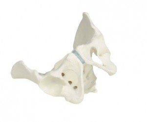
Natural casting of an adult male pelvis.
Wings of ilium, sacrum and flexiblymounted L1 and L2.
The sacrum isremovable and the movements in theiliosacral joint can be demonstrated.
...
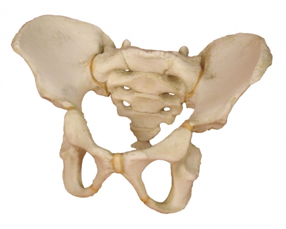
A Life-size male pelvis model with sacral bone showing bone anatomy. The Examples of structures shown in the model are, for example, anterior superior iliac spines, anterior lower bidra spines, sciatic tumors, ischial ...
Actual cast of a real human bony child pelvis.
This model is particularly suitable for explanation of the pelvis development during growth.
The one-part model is not movable.
...
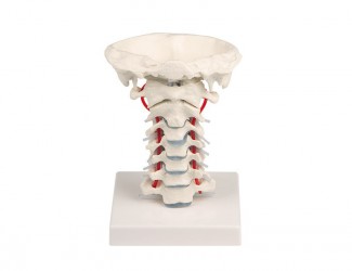
The anatomical model presented here is a life-size cast showing the cervical spine C1 through C7 with the occipital bone flexibly attached, the spinal cord and outgoing spinal nerves. It enables demonstration of ...
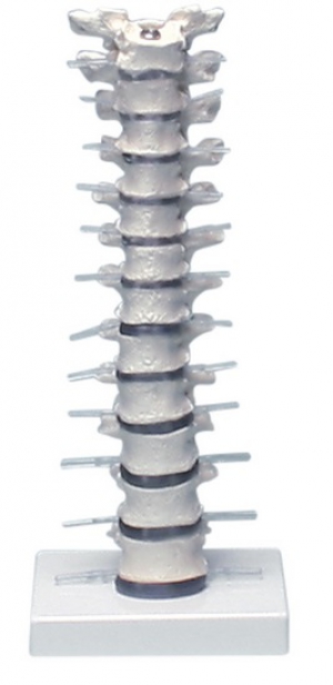
A life-size cast showing the thoracic segment of the human spine (from TH1 oTH12), the spinal cord and the outgoing spinal nerves. Demonstrates intervertebral joints formed by the articular processes of the respective ...
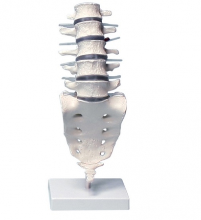
The presented model is a life-size cast of the lumbar spine of the human spine (from L1 to L5) with the sacrum attached in a way that allows showing the mobility of the lumbar spine. The model also presents outgoing ...
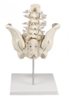
The anatomical model showing the lumbar spine and pelvis. Everything is placed on a stable base. It is possible to remove the model from the base to present detailed anatomical structures. The intervertebral disks ...
