Quality Certyficate
street: Kolejowa 2, 30-805 Cracow


Home
3D anatomy models
Anatomical Models
Custom tools for patient education
Veterinary simulators
Anatomical Charts
Anatomical Table 3D
Medical simulators
Medical Equipment
Type:
 See our profile on Facebook
 Check our profile on Instagram
 Download a PDF file
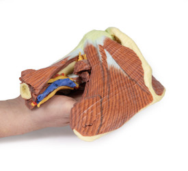
This 3D printed specimen presents a deep dissection of the left shoulder joint, musculature, and associated nerves and vessels of the scapula and proximal humerus (to near midshaft). Anteriorly, the deltoid muscle ...
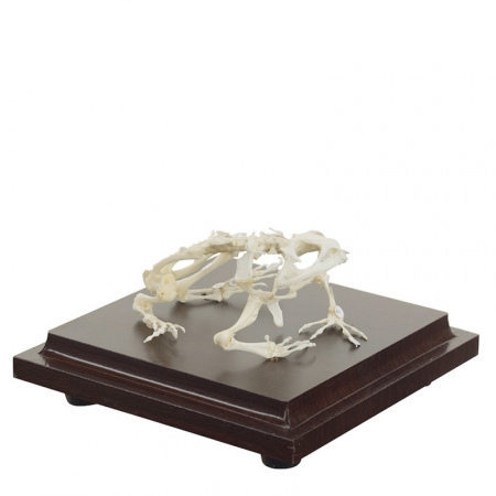
This frog skeleton specimen shows the skeletal structure of this amphibian and the numbered bones are easy to identify
...
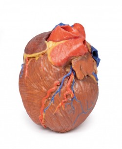
This 3D printed heart specimen preserves superficial cardiac anatomy and the bases of the great vessels. All four chambers (atria and ventricles) are preserved, with the pericardial reflections on the left atrium ...
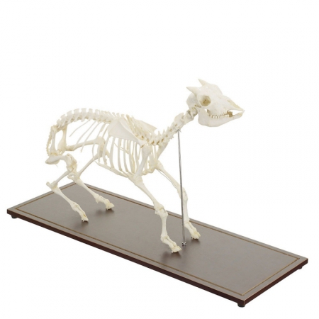
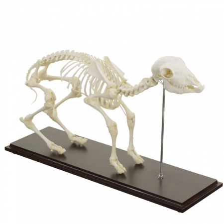
This goat skeleton specimen is used for learning, demonstrating and visualising the animal’s detailed anatomical structure.
Real, life-size goat skeleton
With numbered bones
Delivered with ...
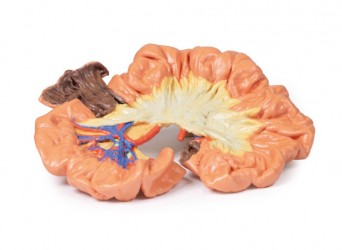
This 3D printed specimen presents a small loop of jejenum and mesentery. A window into the mesentery, fat and visceral peritoneum has been removed to illustrate the arterial arcades in the mesentery (many long ...
This sheep skeleton specimen is suited for visualising the anatomical structures of a sheep and the individual bones are numbered for accurate identification.
Real skeleton of a sheep
Firmly ...
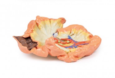
This 3D print displays the orbital contents and its close relations as viewed from the medial perspective when the majority of the lateral wall of the nasal cavity and the intervening ethmoidal sinuses have been ...
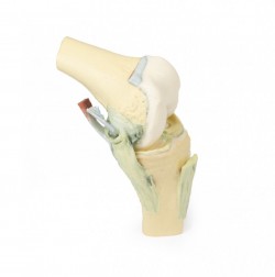
This 3D printed specimen demonstrates the ligaments of the knee joint with the leg in flexion. In the anterior view, with the patella and part of the patellar ligament removed, the medial and lateral menisci and ...
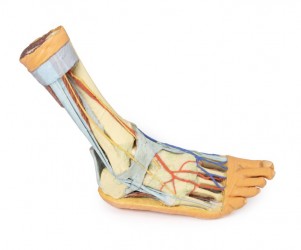
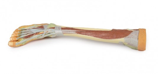
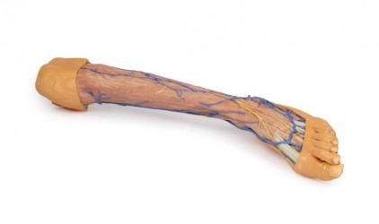
This 3D printed specimen presents both superficial and deep structures of a right distal leg and foot. Proximally, the posterior compartment of the leg has been dissected to remove the triceps surae muscles and ...
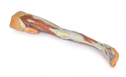
This 3D printed specimen preserves the distal thigh and proximal leg, dissected posteriorly to demonstrate the contents of the popliteal fossa and surrounding region. The proximal cross-section demonstrates the ...
This 3D printed specimen demonstrates a small loop of ileum and mesentery. A window into the mesentery has been dissected (removing fat and visceral peritoneum) to show arterial arcades in the mesentery (many short ...
High Quality anatomical chart, made of 200µ plastic foil, size appr. 70x100cm, incl. metal edging with hanger cord. Nomenclature in German, Latin and English. ...
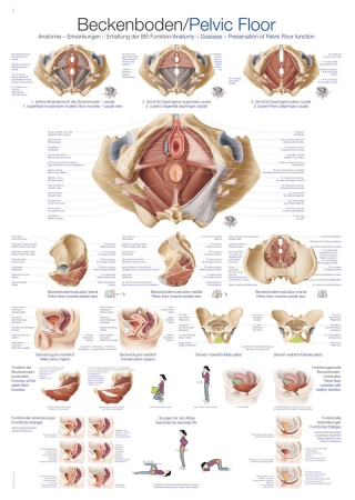
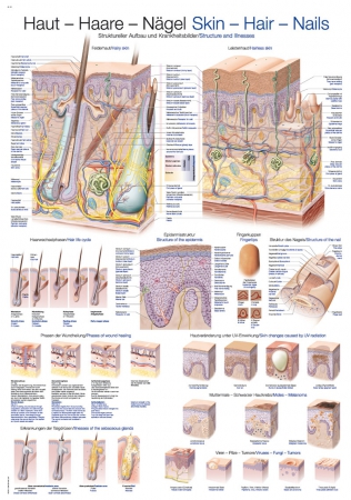
High Quality anatomical chart, made of 200µ plastic foil, size appr. 70x100cm, incl. metal edging with hanger cord. Nomenclature in German and English. ...
High Quality anatomical chart, made of 200µ plastic foil, size appr. 70x100cm, incl. metal edging with hanger cord. Nomenclature in German and English. ...
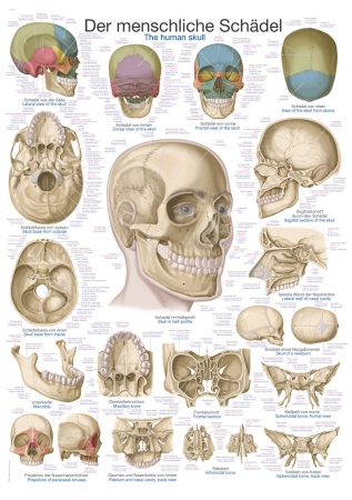
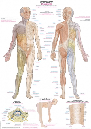
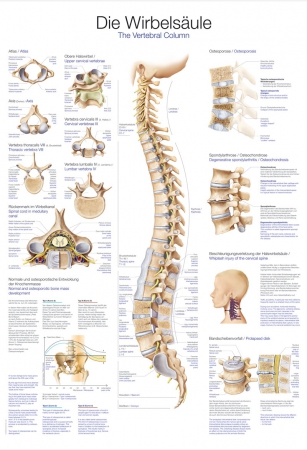
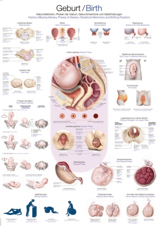
High Quality anatomical chart, made of 200µ plastic foil, size appr. 70x100cm, incl. metal edging with hanger cord. Nomenclature in German, Latin and English. ...
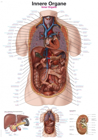
High Quality anatomical chart, made of 200µ plastic foil, size appr. 70x100cm, incl. metal edging with hanger cord. Nomenclature in German, Latin and English. ...
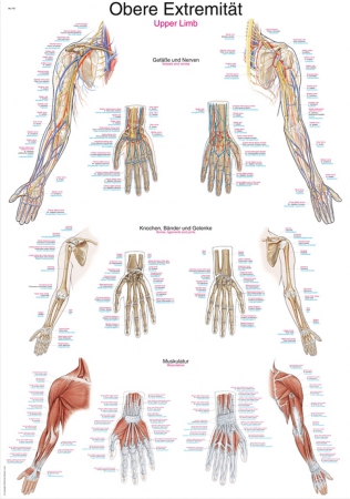
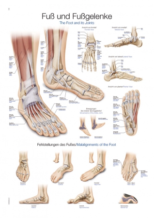
High Quality anatomical chart, made of 200µ plastic foil, size appr. 70x100cm, incl. metal edging with hanger cord. Nomenclature in German and English. ...
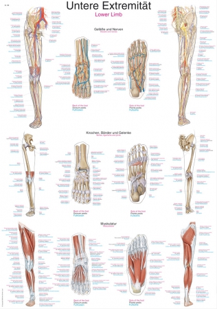
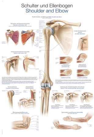
High Quality anatomical chart, made of 200µ plastic foil, size appr. 70x100cm, incl. metal edging with hanger cord. Nomenclature in German and English. ...
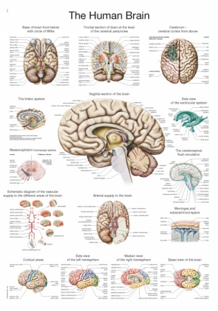
High Quality anatomical chart, made of 200µ plastic foil, size appr. 70x100cm, incl. metal edging with hanger cord. Nomenclature in English. ...
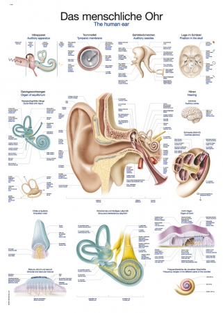
High Quality anatomical chart, made of 200µ plastic foil, size appr. 70x100cm, incl. metal edging with hanger cord. Nomenclature in German, Latin and English. ...
High Quality anatomical chart, made of 200µ plastic foil, size appr. 70x100cm, incl. metal edging with hanger cord. Nomenclature in German and English. ...
High Quality anatomical chart, made of 200µ plastic foil, size appr. 70x100cm, incl. metal edging with hanger cord. Nomenclature in German and English. ...
High Quality anatomical chart, made of 200µ plastic foil, size appr. 70x100cm, incl. metal edging with hanger cord. Nomenclature in German and English. ...
High Quality anatomical chart, made of 200µ plastic foil, size appr. 70x100cm, incl. metal edging with hanger cord. Nomenclature in German and English. ...
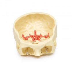
This 3D printed specimen demonstrates the intracranial arteries that supply the brain relative to the inferior portions of the viscero- and neurocranium. This print was created by careful segmentation of ...
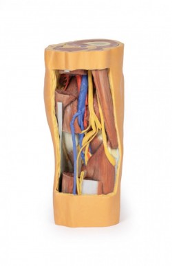
This 3D printed specimen consists of a right partial lower limb sectioned just proximal to the knee joint and complete through a partially dissected foot exposing the structures on the dorsum. In the proximal cross ...
Lower Limb:
sectioned proximally near midthigh and continuous to the partially dissected foot. The transverse section through the thigh exposes the neurovascular structures of the anterior, medial and posterior ...
A professional model of the head of an adult human, designed to perform MRI, CT and ultrasound imagining.
These phantom are used for medical imaging (both static and flow) and for treatment planning for ...
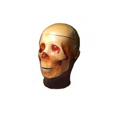
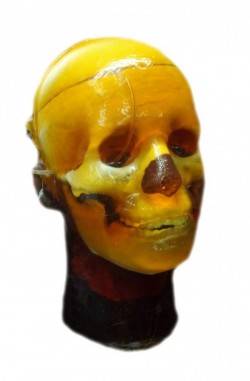
A professional phantom head designed for medical imaging of foreign static objects, such as small bones, shards, spheres and other metal or non-metallic objects. It was designed based on a medium-sized man's head. Made ...
This 3D printed specimen presents a superficial dissection of a left lower limb, from just proximal to the knee joint to a complete foot. The skin and superficial fascia have been removed to display the superficial ...

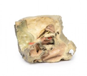
This 3D model of the head and neck represents a specimen sectioned just off the midsagittal plane to retain some midline anatomical structures (e.g., the falx cerebri, the septum pellucidum, the nasal septum) that are ...
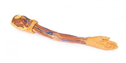
This 3D print demonstrates a superficial dissection of a left upper limb from the blade of the scapula to the hand. The skin, superficial and deep fascia has been removed from most of the limb except over the dorsum of ...
