Quality Certyficate
street: Kolejowa 2, 30-805 Cracow


Home
3D anatomy models
Anatomical Models
Custom tools for patient education
Veterinary simulators
Anatomical Charts
Anatomical Table 3D
Medical simulators
Medical Equipment
Type:
 See our profile on Facebook
 Check our profile on Instagram
 Download a PDF file
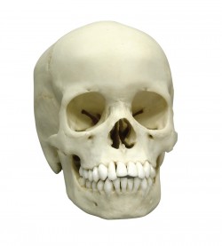
This skull offers an excellent example of an adolescent. With the exception of the wisdom teeth, all permanent teeth are fully erupted, and no deciduous dentition remains. The apices of the canines, premolars, and ...
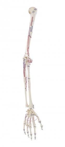
Life-size bone model of the upper limb with muscle attachment markings. It shows the following bones:
humerus (humerus),
ulna (ulna),
radius ...
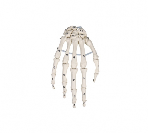
True to life casting of a skeleton of the human hand. All hand bones are individually mobile-mounted on wire. With additional numbering of the individual hand bones. ...
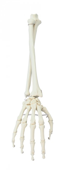
Life-size bone model of the hand and forearm. It presents all the bones of the hands: wrist, metacarpus, phalanges. Has the ability to perform translatorial and anatomical movements between individual bones. ...
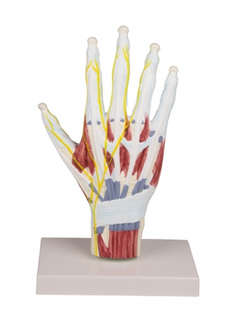
Life-size anatomical model of the hand. It presents hand-made muscles of the hands, ligaments, nerves, bones and tendons. Made on the basis of. There is a possibility remove the model from the base.
Additional ...
Natural casting of a human leg. Can be dismantled into femur, tibia, fibula and foot.
...

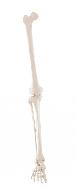
Natural casting of a human leg.
Can be dismantled into femur, tibia, fibula and foot. With marking of the muscle origins and insertion points.
...
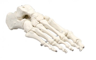
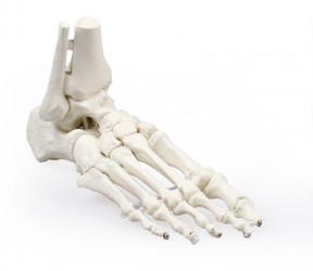
Natural casting of a human foot. All bones mobile mounted on wire.
Without stand.
...
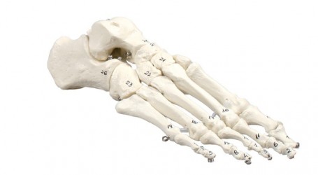
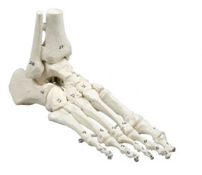
Natural casting of a human foot. All bones mobile mounted on wire.
Model as KD6050, but with bone numbering.
Without stand.
...
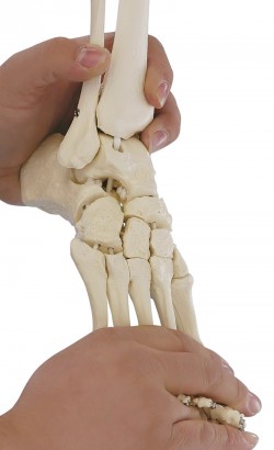
Natural casting of a human foot mounted on wire.
With tibia and fibula insertion.
Without stand.
...
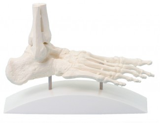
Natural casting of a human foot mobile mounted on wire.
With tibia and fibula insertion.
Without stand.
...
A life-size elastic model of the human foot with fragments of the lower leg bones. Individual bones have been connected with a special elastic band, which makes this model very mobile. It is a feature that distinguishes ...
Natural one-piece casting of a human foot.
Representation of all structures and anatomical details.
Particularly economically priced.
...
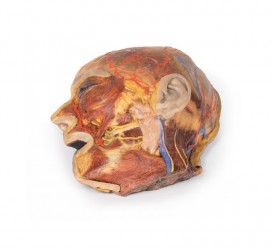
This 3D model presents the superficial anatomy of the face and head, and compliments the superficial facial anatomy of our HW 44 model with a more expanded dissection across the scalp and occipital regions. The ...
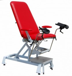
The S02 gynecological chair has a stable steel structure covered with powder paint, the additional durability of the chair is ensured by the use of stainless steel in the movable parts of the chair. The top consists of ...
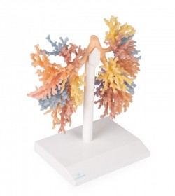
The highly detailed 3D model of the bronchial tree shows the airways from the trachea, spurs trachea and the entire right and left bronchial tree to the bronchial level of the tertiary lobes. Each lobe bronchus system ...
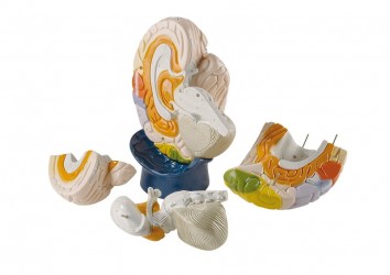
This 3D printed model captures a dissection in which the calvaria and cerebrum have been removed to expose the floors of the anterior and middle cranial fossae. The midbrain has been sectioned at the level of the ...
This 3D printed specimen shows the orbit from the lateral perspective when the bony lateral wall and part of the calvaria of the skull have been removed. The frontal and temporal lobes of the brain are exposed. In ...
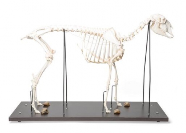
Prepared, real skeleton of a domestic sheep consisting of approximately 215 individual bones, which are rigidly connected to each other. It represents a typical example of the order of even-toed ungulates. You can ...
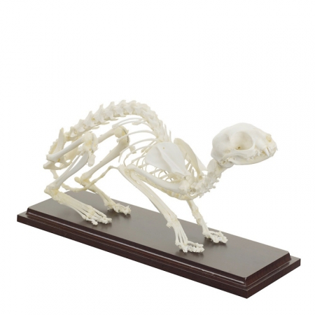
A skeleton model of a cat on a wooden base. It is an excellent didactic aid, it is used for demonstration purposes.
Additional features:
dimensions: 33x9x16cm
has individual bones ...
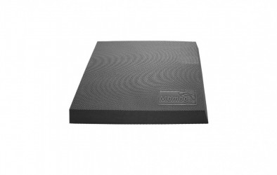
Balance Pad for exercises to strengthen the feet, ankles and leg muscles. Perfect for balance training and sensorimotor tasks.
Specification:
dimensions: 47x39x6cm
weight: 07,kg
color: ...
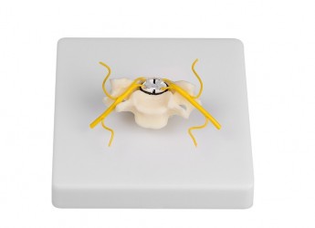
Individual cervical vertebra with cross sectional representation of the spinal cord. White and grey spinal cord substance and the spinal nerves shown. On baseboard. ...
This unique model shows an enlarged human neuron. The axon shows a healthy myelin sheath and three stages of myelin sheaths affected by multiple sclerosis. The neuron can be removed from the base for closer ...
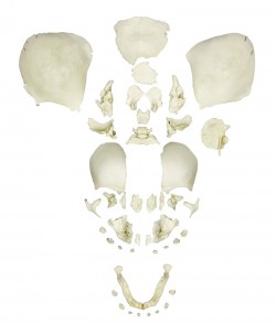
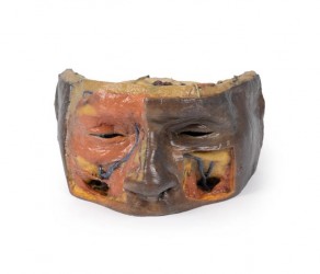
This disarticulated skull is from a full term (10 lunar months) fetus. It was part of a medical examiner‘s comparative pathology collection before going to the Maxwell Museum and is remarkable in its completeness. It ...
Model twice enlarged, divided into 4 parts. The frontal lobes and brainstem are removed.
specifications:
dimensions: 36cm x 28cm x 20cm
advanced painting
delivered based on
...
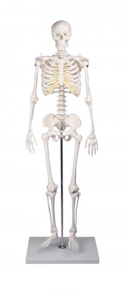
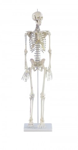
The model shows a twice-reduced human skeleton. Skulls, arms and legs are removed. In addition, the skull can be divided into three parts.
Specifications:
dimensions: approx. 84 cm high (without the base)
...
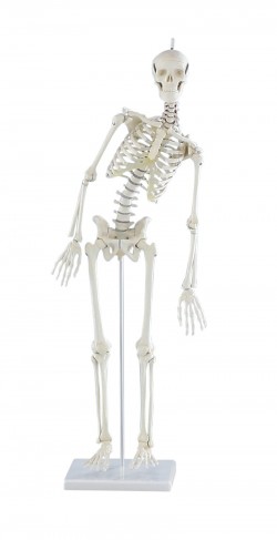
The model shows a twice-reduced human skeleton. The arms and legs are fully movable; shoulders, hips, knees and ankles are mounted as sliding joints to allow natural movement. The three-part skull and the arms and legs ...
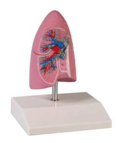
Right lung model doubled. Includes:
bronchi,
arteries,
veins.
Specifications:
dimensions: 11cm x 7cm x 7cm,
weight: approx. 0.2 kg,
advanced painting,
delivered ...
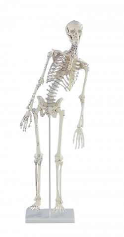
The model shows a twice-reduced human skeleton. The arms and legs are fully movable, the arms, hips, knees and upper ankles are designed as sliding joints, so you can show all natural movements. The three-part skull, ...
The model shows a twice-reduced human skeleton. The arms and legs are fully movable, the arms, hips, knees and upper ankles are designed as sliding joints, so you can show all natural movements. The three-part skull, ...
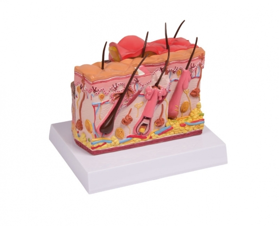
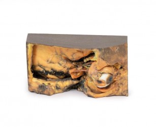
Enlarged 2-sided skin cross-section. The burn side of the model shows indication of 1st, 2nd and 3rd degree burns. The reverse side illustrates normal skin anatomy. With stand.
Size: 15 x 10 x 7 cm ...
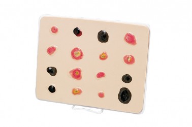
Continued exposure to sunlight can cause damage to skin cells. If the damaged cells do not die or repair themselves, they degenerate and visible skin cancer develops. The Skin Cancer Trainer has been developed to ...
