Quality Certyficate
street: Kolejowa 2, 30-805 Cracow


Home
3D anatomy models
Anatomical Models
Custom tools for patient education
Veterinary simulators
Anatomical Charts
Anatomical Table 3D
Medical simulators
Medical Equipment
Type:
 See our profile on Facebook
 Check our profile on Instagram
 Download a PDF file
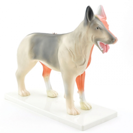
The disassemblable canine model comprises two halves, one of which shows the outside of the dog, depicting the fur and mucous membranes, while the other half of the model shows the dog without skin, ...
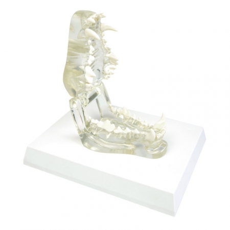
Product information:
The transparent canine jaw model visualises the healthy masticatory apparatus of an adult dog.
Transparent model of upper and lower jaw of an adult dog
Upper and lower jaws can ...
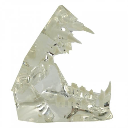
Product information:
The transparent feline jaw provides a detailed visualisation of the healthy dentition of a cat.
The See-Through Cat Dentition Shows:
Incisors
Canines
Premolars
...
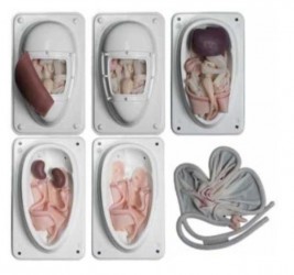
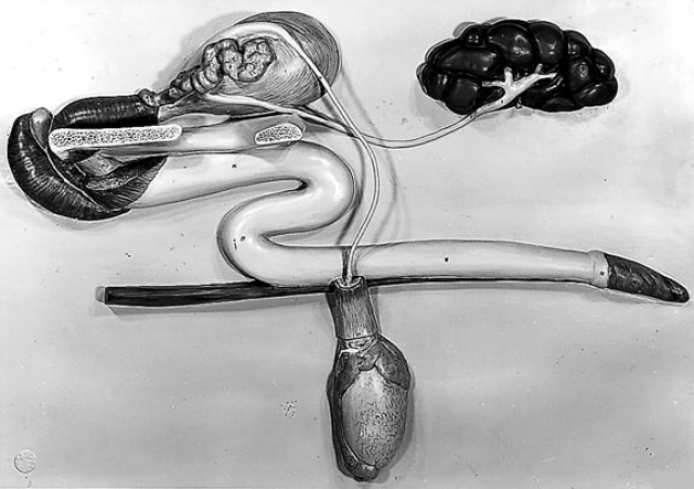
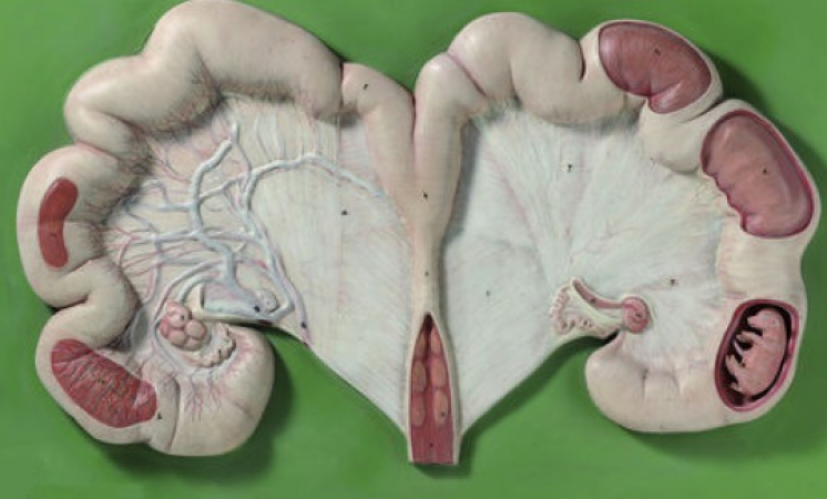
The simulator intended for learning and training dog sterilization.
Main features:
Replaceable uterus with ovaries, broad ligament and suspensory ligaments
Large and small intestine with mesentary and ...
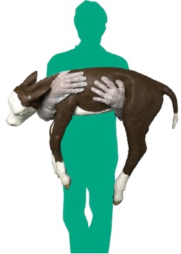
A realistic model of a dystocia calf.
Main features:
Fully articulated steel skeleton, with ribcage, vertebrae, pelvis and spine
Realistic movement; durable and flexible skin and tail
Skull with ...
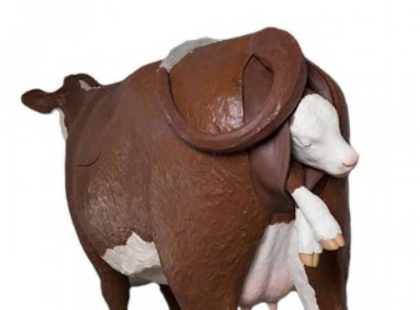
Dystocia simulator for the Hereford breed.
Main features:
Steel reinforced epoxy/fiberglass construction, with water resistant components throughout for ease of cleaning.
1.36m at the shoulder and over ...
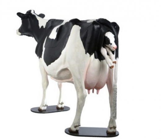
Dystocia simulator for the Holstein breed
Main features:
Steel reinforced epoxy/fiberglass construction, with water resistant components throughout for ease of cleaning.
1.57m at the shoulder and over 2.80m ...
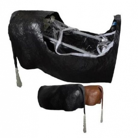
Realistic performed, modeled on the Hereford breed, a compact dystocia simulator.
Main features:
Steel reinforced epoxy/fiberglass construction, with water resistant components throughout for ease of cleaning.
...
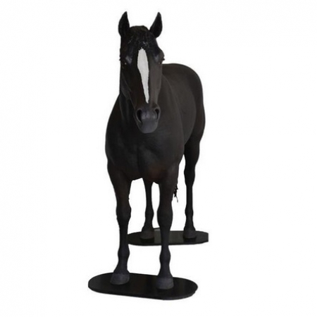
A realistic palpation horse simulator in natural size. It allows performing venipuncture and intramuscular injection.The simulator comes equipped with inflating latex intestines to familiarize students with the ...
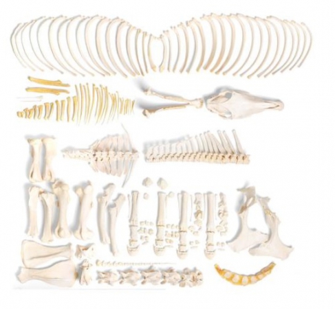
Complete and non-assembled skeleton bones without pre-drilling. Ideal for demonstrating the typical bone structure and anatomy. Small bones can be loose. Does not include building instructions. ...
This impressive specimen of a genuine cow skeleton comes mounted on a wooden base for easy display in the classroom. There is no better way to study the anatomy Bos taurus, the domestic cow, then with this cow skeleton. ...
Complete and non-assembled head without pre-drilling. Ideal for demonstrating the typical bone structure and anatomy. Small bones can be loose. Does not include building instructions. ...
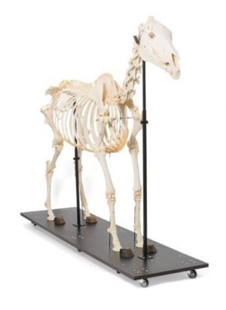
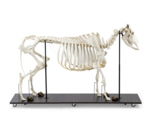
The real bone specimen of an adult horse is composed of approximately 252 individual bones. It represents a typical example of the order of odd-toed ungulates. There are only minimal di?erences in body plan between ...
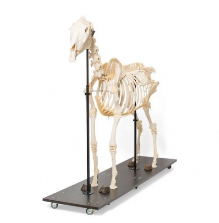
The real bone specimen of an adult horse is composed of approximately 252 individual bones. It represents a typical example of the order of odd-toed ungulates. The skeleton is mounted on a moveable base ...
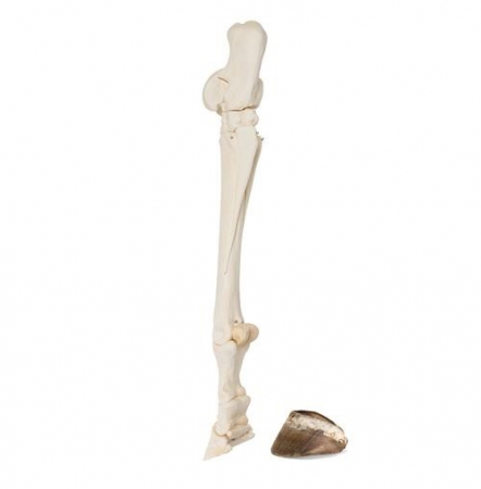
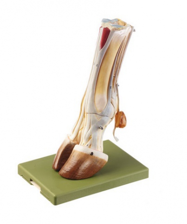
Prepared, real, hind horse foot up to the tarsal joint. The individual bones are rigidly connected to each other. The hoof capsule is supplied separately with the foot.
Horse (Equus ferus ...
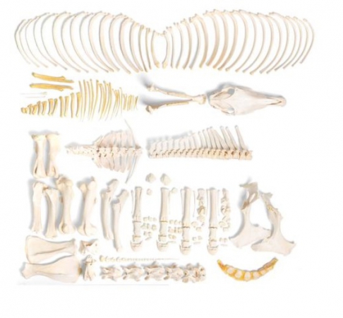
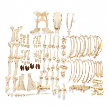
Complete and non-assembled skeleton bones without pre-drilling. Ideal for demonstrating the typical bone structure and anatomy. Small bones can be loose. Does not include building instructions.
Horse (Equus ...
Complete and non-assembled skeleton bones without pre-drilling. Ideal for demonstrating the typical bone structure and anatomy. Small bones can be loose. Does not include building instructions.
Horse (Equus ...
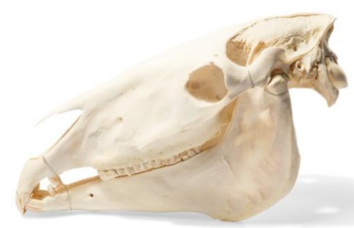
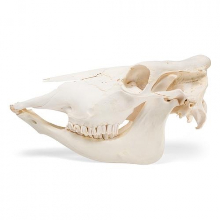
Bone specimen of a horse skull consisting of approximately 37 individual bones, which are rigidly connected to each other. All the teeth are firmly attached to the jaws.
Length: Approx. 60 cm
Horse (Equus ...
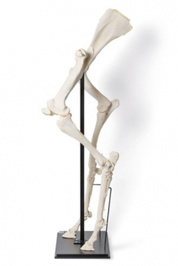
Real bone specimens from an adult horse. Each front leg up to and including the shoulder blade and each hind leg up to the hip joint. Rigidly mounted on a base plate
Horse (Equus ferus ...
Product information:
Natural size. In one piece. Mounted on a board.
...
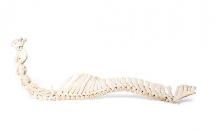
Real horse spine with cervical, thoracic, lumbar, and sacrum. Flexibly mounted.
Horse (Equus ferus caballus)
Taxonomy:
Class: Mammals
Order: Odd-toed ungulates
Family: Horses
Diet: Herbivore
Size: Approx. ...
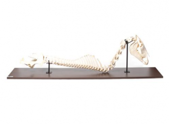
Real horse spine with cervical, thoracic, lumbar, sacrum, and head. Rigidly mounted.
Horse (Equus ferus caballus)
Taxonomy:
Class: Mammals
Order: Odd-toed ungulates
Family: Horses
Diet: Herbivore
Size: ...
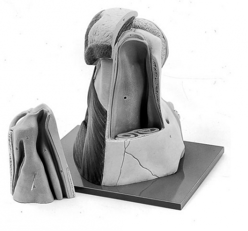
Product information: Nose of Cow
Natural size, modelled from a natural preparation. The model shows the exact anatomical structure, the bony surround, the muscles, the nasal cartilages, glands and moist part of ...
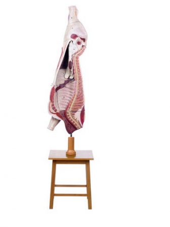
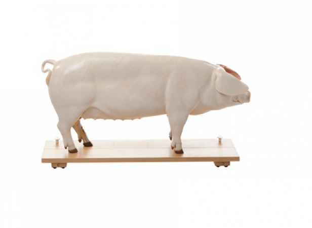
Product information: Model of the Carcass of a Bullock
1/2 natural size. Produced in collaboration with the Bavarian Institute for Animal-Breeding in Grub near Munich. The model shows the left half of the ...
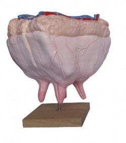
The cow udder model separates into 4 parts in sagittal and vertical section, showing the arteries, veins and milk passages and the four glandular regions. Removable. On a stand with wooden base.
Dimension: ...
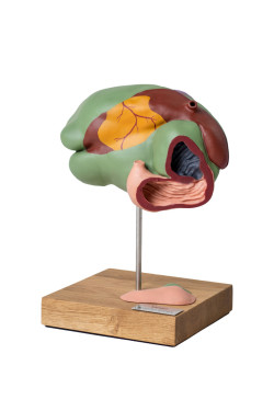
Rumen and reticulum can be divided into two halves to show the relief of the mucous membrane of the stomach. Omasum and abomasum can be opened up. Separates into 3 parts.
...
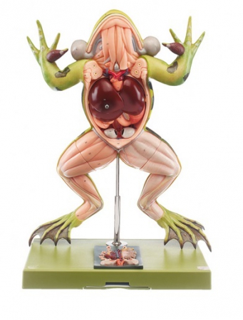
4 times enlarged male frog model with spread legs and inflated voice bags. The view from the back shows typical anatomical elements (marked with colors and numbered). The liver and the gastro-intestinal tract ...
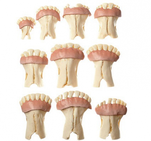
Product information:
Natural casts of the lower jaw showing ten different stages of growth: 14 days, 1 year, 1 1/2 years, 2 years, 3 years, 4 years, 5 years, 9 years, 14 years, and 18 years. In one piece. ...
Product information: Cow's Hoof
Left front foot of the cow, cast from natural specimen. Separates into 6 parts. On a green base.
...
Right side shows the skin, the other side shows the muscular system. The model is mounted on a base which can be pulled out and separates into two halves medially. The left half of the head showing the muscular system, ...
Product information:
Natural size, in one piece. Mounted on a board.
...
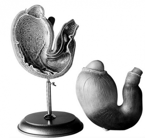
Product information: Stomach of the Pig
Natural size. Can be opened to show the relief of the folds of the mucous membrane. Separates into 2 parts. On a stand and base.
...
