Quality Certyficate
street: Kolejowa 2, 30-805 Cracow


Home
3D anatomy models
Anatomical Models
Custom tools for patient education
Veterinary simulators
Anatomical Charts
Anatomical Table 3D
Medical simulators
Medical Equipment
Type:
 See our profile on Facebook
 Check our profile on Instagram
 Download a PDF file
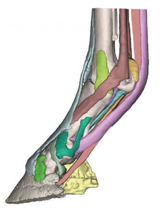
Derived from CT and MR co-registered data, anatomically accurate and lifesize. 3D printed in full color, each anatomical component is individually colored. Hoof capsule is available separately and anchors the distal ...
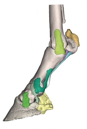
Derived from CT and MR co-registered data, anatomically accurate and lifesize. 3D printed in full color, each anatomical component is individually colored. Hoof capsule is available separately and anchors the distal ...
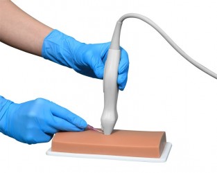
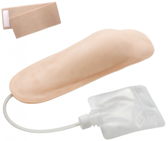
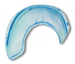
The is an excellent training phantom for the introduction and improvement of the techniques and psychomotor skills associated with successful ultrasound-guided vascular accessing. The unique material is flesh ...
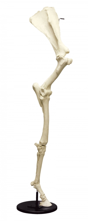
We include the scapula, humerus, forearm (fused radius and ulna), cannon bone and foot. The foot consists of the long pastern bone (or first phalanx), the short pastern bone (or second phalanx) and the coffin bone ...
This ultrasound model shows a left, life-size human knee with the following structures:
Femur, tibia and patella bones. Quadriceps tendon, patellar ligament, infrapatellar fat pad, synovium, prepatellar bursa, ...

This ultrasound model shows a life-size human shoulder with the following structures:
Humerus, scapula, clavicle, acromioclavicular ligament and biceps ligament.
The shoulder can be punctured with 27 gauge ...
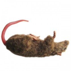
The Squeekums Rat manikin allows students, lab technicians, and handlers to learn how to confidently handle and safely manage lab rodents. The tail of this amazing rodent model can be conveniently detached and ...
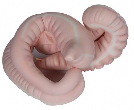
A realistic model imitating the horse's gastrointestinal tract. Anatomically correct.
The model consists of 5 pumped components (Each component above is divided into batches separately to allow individual ...
We hope to contribute to a reduction in the number of experimental animals used, and is used as an alternative to animal experiments in medical, pharmaceutical, and veterinary education and the education for ...
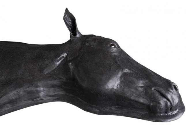
The (in black) is the leading and most comprehensive equine simulator available. It has been designed to advance a comprehensive set of educational goals for the following procedures:
- Intramuscular ...
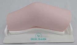
With this model you practice directly at the patient. It can be attached to a simulated patient or a nursing doll. With excellent haptics it is the ideal tool for teaching the i.m. injection technique at the upper ...
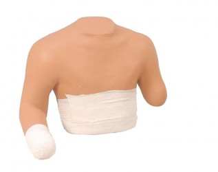
Upper torso with amputation stumps for bandaging training.
*Life size ...
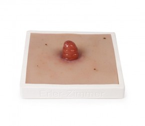
This moulage shows an ileostomy, the second most common type of ostomy. The terminal ileostoma is usually found on the right side of the body and shows the small intestine sewn in above skin level.
Addictional ...
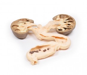
Clinical History
A 49-year old male presents with a 6-week history of malaise, urinary frequency and haematuria for 6 weeks. Further questioning revealed intermittent left flank pain. Abdominal ultrasound showed ...
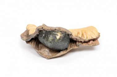
Clinical History
A 54-year-old man presented to hospital with 12 hours of severe colicky pain, nausea and vomiting. On history, he was noted to have had a 3-year history of intermittent right subcostal pain for ...
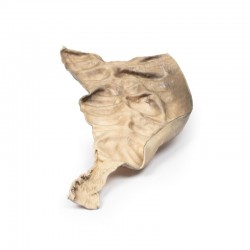
Clinical History
A 5-year old male presents with a history of constipation since birth. A barium enema showed a constricted rectum with a dilated sigmoid colon. Surgical resection of constricted section of bowl ...
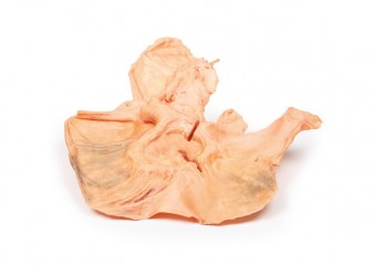
Clinical History
An 82-year old female presents with an episode of melena (dark tarry faeces). She had a 6-month history of dyspepsia and nausea. Recently she had noted weight loss and early satiety. Soon after ...
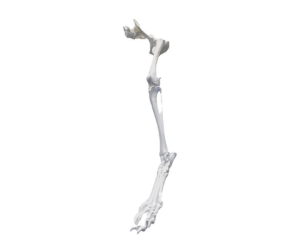
Anatomically precise 3D printed dog bones.
3D printed bones accurately show important osteological features of the hind limb, from the hip down to the digits, enabling students to learn and identify the ...
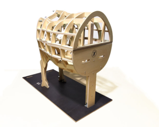
Holsim's is a flat-pack, transportable training mannequin for classroom teaching of calving techniques. This interlocking birthing cow is made from premium grade plywood pieces that slot and bolt together for easy ...
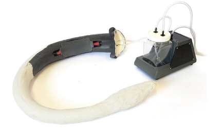
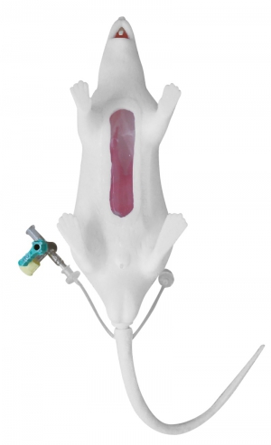
Model of a cow tail with artificial blood pump and lifelike coccygeal vein for practicing blood taking techniques.
Anatomically Correct
Vertibrae and vein positioning have been replicated from ...
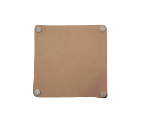
A training pad for practising surgical stitching.
Holsim Suture Training Pads allow students to maximise surgical stitching techniques. Tutors can teach their students needle placement, stitching patterns, knot ...
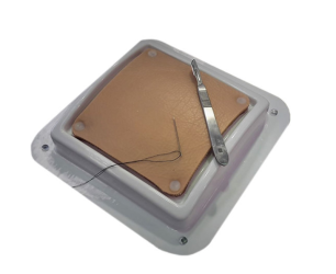
Holsim Suture Training Pads allow students to maximise surgical stitching techniques. Tutors can teach their students needle placement, stitching patterns, knot tying, superficial and surgical closures. Suture Training ...
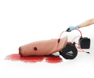
The P103 is a medical skill trainer for hemorrhage control on the lower extremity with an extremely realistic wound and bleeding simulation. It is the ideal tool to improve pre-hospital patient care training with ...
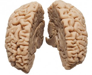
This model of the brain was formed from a real human brain. Special materials and an elaborate manufacturing process allow for a previously unknown level of detail in recreating structures in anatomy lessons. The left ...
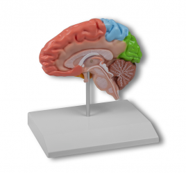
This model shows the main landmarks of the brain in well-represented colors. It is painted so that the four lobes of the brain can be distinguished: the frontal and lateral lobes, and the temporal and occipital lobes. ...
This model represents a cerebral falx. It shows the orifices of the sternal veins, arachnoid villi, superior and inferior sagittal sinus and straight sinus. ...
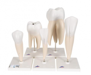
This classic tooth series shows 5 representative types of adult dentition individually mounted on removable stands:
2-part lower incisor with longitudinal section
2-part lower canine with longitudinal section
Lower ...
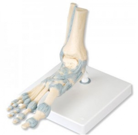
This detailed foot skeleton model displays numerous important ligaments and tendons including the Achilles and peroneus longus tendons of the ankle. The foot skeleton consists of the foot bone and lower portions of the ...
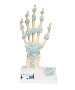
This 3 part hand skeleton model shows the anatomical detail of the ligaments and tendons found in the hand, wrist, and lower forearm. The interosseous membrane between the radius and ulna is shown along with the bones ...
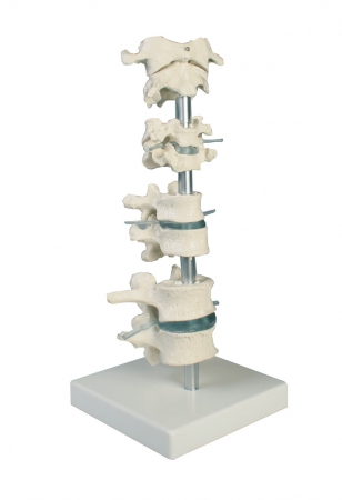
The model presents 8 individual vertebrae of different sections of the spine
The model includes:
2 lumbar vertebrae,
2 thoracic vertebrae,
2 cervical vertebrae
atlas and axis
All structures, ...
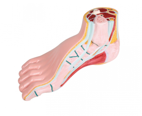
This life size model of a right human foot is medially opened and shows bones, joints, ligaments, fatty tissue and muscles. The start of lower leg is also represented as cross-section and shows tibia and fibula as ...
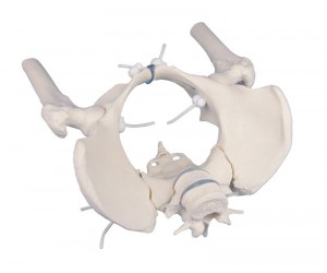
A life-size cast of the female pelvis. It has the ability to disassemble the sacrum and demonstrate mobility in the sacroiliac joint. The model also has two lower lumbar vertebrae. Fragments of the femurs move ...
