Quality Certyficate
street: Kolejowa 2, 30-805 Cracow


Home
3D anatomy models
Anatomical Models
Custom tools for patient education
Veterinary simulators
Anatomical Charts
Anatomical Table 3D
Medical simulators
Medical Equipment
Type:
 See our profile on Facebook
 Check our profile on Instagram
 Download a PDF file
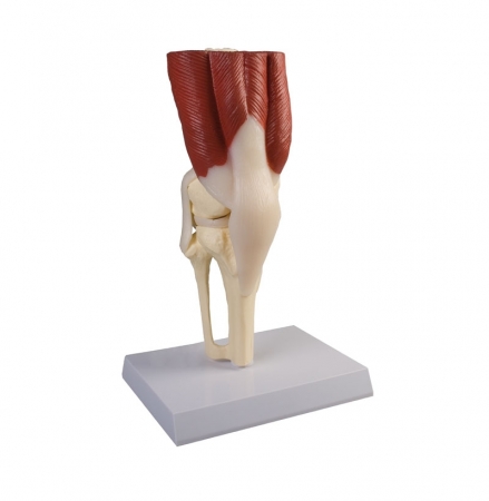
Human knee joint in life size with all important muscles and ligaments (collateral ligaments, meniscus, crucial ligaments, patellar tendon).
The joint is not movable.With educational card German/English.
On ...
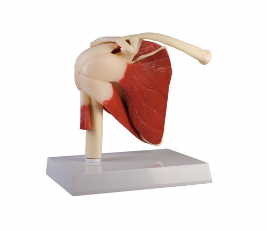
Human shoulder joint in life size with rotator cuff (Supraspinatus, infraspinatus, teres minor, teres major and subscapularis muscle) as well as the biceps brachii tendon. The joint has a limited movability.
With ...
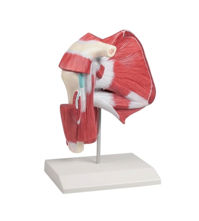
This model illustrates in great details the muscles, ligament and bones of the shoulder.
Through different muscles section it is possible to observe the profound musculature as far as to the bone.
...
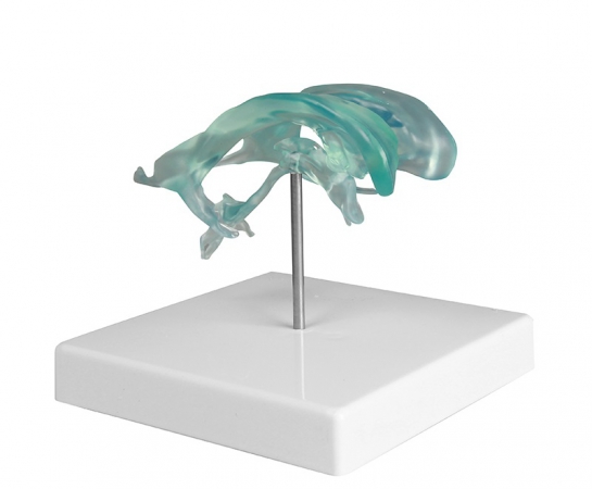
Life size model of the human lateral ventricles, cerebral aqueduct, as well as the 3rd and 4th ventricle. With removable stand. ...
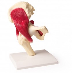
Human hip joint in life size with all important muscles and ligaments. The joint has a limited movability. With educational card German/English.
On Stand.
Size: 13 x 13 x 24 cm, weight: 0.7 kg ...
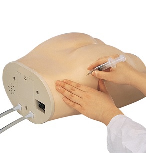
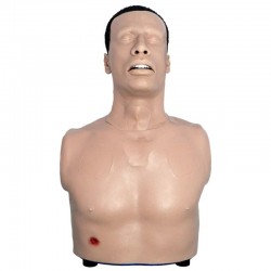
This highly realistic model consists of a special plastic that gives the user the feeling of working on an actual human patient. All anatomical structures are present and palpable. The injection sites are constructed ...
Main Features
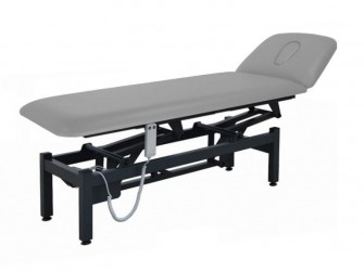
2-section therapy couch
simple and stable steel frame (superior stability)
Electrical height adjustment (Hand switch)
Gas-spring head section adjustment
A wide range of ...

The presented model was made in such a way as to present the life-size human head with the brain, the position of the arterial and venous vessels of the head, Willis arterial circle, cranial nerves and brainstem. The ...
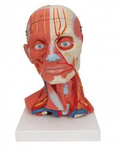
Detailed head and neck model on the base. It presents the muscles of the head, back, neck, face, blood vessels and news. The model can be disassembled for a total of 5 parts. They are: three-part brain, skull cover, ...
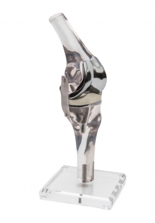
This life size knee model is made of transparent plastic and shows graphically the position and function of a knee endoprosthesis.
The reproduced prosthesis can be removed and replaced from the model, allowing to ...
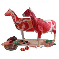
A professional, fully colorful, didactic horse model on the base. It has the ability to divide into 12 separate parts of the body and organs. The model shows the muscular structure of the goat on one side, on the ...
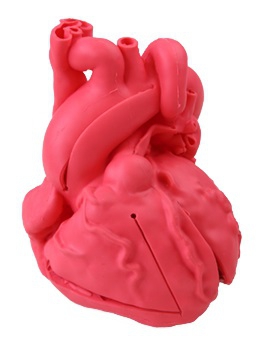
2-times life size heart model is based on CT scan data of a healthy, adult male and is anatomically correct inside and out. Reproduced sections: (external & luminal surfaces) right atrium, left atrium / right ...
The TacMed Simulation APL-C can be used during the crawl phase of training. It trains responders to perform life-saving tasks such as maintaining a patient’s airway, needle ...
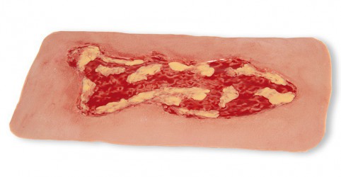
This self-adhesive wound represents a venous leg ulcer, a substance defect in the tissue of the lower leg as a result of a chronic venous insufficiency. The shown exudation phase represents the most common ulcus ...
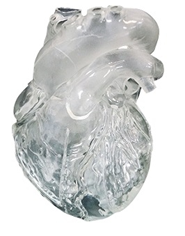
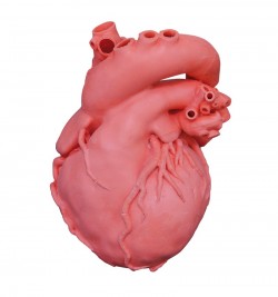
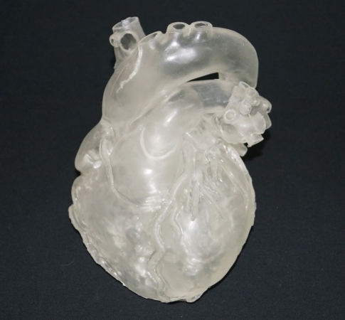
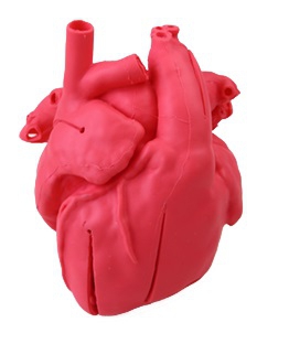
This world-wide unique model is based on CT scan data of a healthy, adult male and is anatomically correct inside and outside. The heart is made of soft and lifelike material and translucent. It is pre-cut at ...
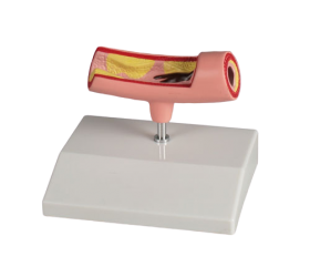
Longitudinal section of an artery with constriction caused by plaque adsorption and a blockage caused by a thrombus.
specifications:
dimensions: 10cm x 4cm x 4cm
weight: approx. 0.1 kg
advanced ...
The pediatric congenital heart disease model is based on actual CT data, and is a precisely produced, urethane based, soft model. This product was created with the intention of being a support tool for doctors ...
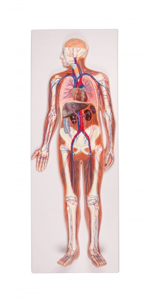
Model of human circulatory system on base basis. Ideal for demonstrating and understanding the mechanism of blood circulation in the human body. The model clearly shows lung circulation, the heart with ventricles, ...
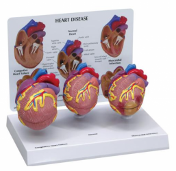
3-Mini Heart Model set. Normal heart anatomy; myocardial infarction heart demonstrating a partially healed, thinned, and discolored infected area. Including a thrombus in the apex of the heart; and a heart demonstrating ...
The pediatric congenital heart disease model is based on actual CT data, and is a precisely produced, urethane based, soft model. This product was created with the intention of being a support tool for doctors ...
This 2-times life size heart model is based on CT scan data of a healthy, adult male and is anatomically correct inside and out. Reproduced sections: (external & luminal surfaces) right atrium, left atrium / ...
The pediatric congenital heart disease model is based on actual CT data, and is a precisely produced, urethane based, soft model. This product was created with the intention of being a support tool for doctors ...
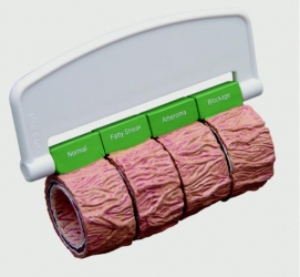
Four-stage cross-section of an artery demonstrating atherosclerosis in which the narrowing of the artery is due to a build up of fatty tissue (cholesterol) and plaque. Four stages: normal artery, fatty streak, fibrous ...
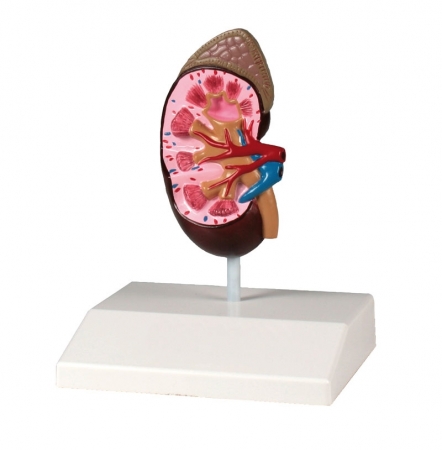
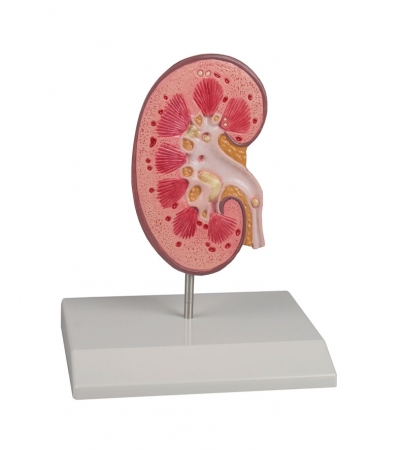
This model of a human kidney in almost life size shows hand painted details of renal pelvis, renal medulla, renal calyx, renal cortex, renal artery and vein, ureter and adrenal gland. With educational card and ...
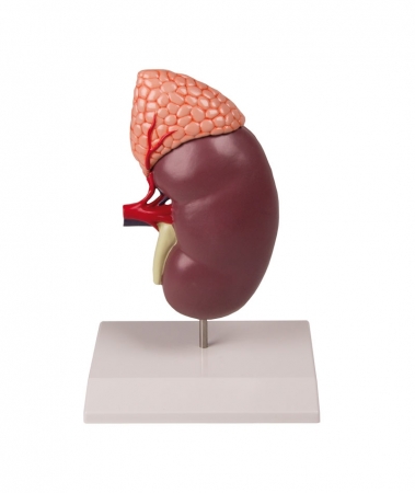
This 2-part model shows the human kidney in about double life size. It shows internal structures including cortex, medulla, pyramids, calyces, renal pelvis, ureter and origins of the renal artery and vein. The front ...
This model is designed to inform patients about urinary stones (urolithiasis) and kidney stones (nephrolithiasis). A right kidney in natural size is opened to show the internal structures. The renal pelvis, the renal ...
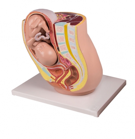
This life size model depicts the female human pelvis in median section with a fetus in the 32nd week of pregnancy. The fetus is in the normal presentation and position. The model depicts graphically the position and ...
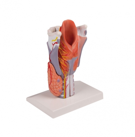
Enlarged larynx model consisting of five parts and in longitudinal section showing the structure of the larynx. These are: hyoid bone; cartilage; ligaments; muscles; blood vessels; nerves; ...
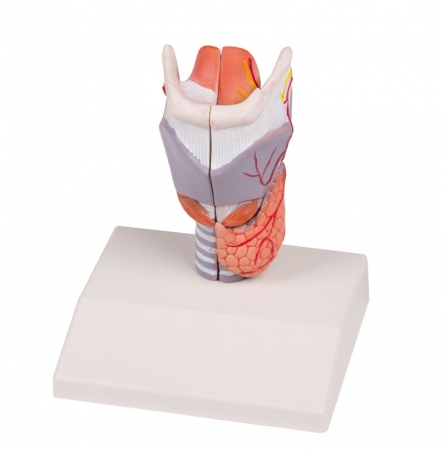
A life-size larynx model that can be broken down into 2 parts. The model presents the following structures: hyoid bone cartilage muscle tendons, dishes nerves thyroid.
Additional ...

The mid-section of the nose and the sense of smell at 4 times magnification. It presents in a detailed way the nasal septum together with nerves and vessels (right side), individual structures of the inner nasal cavity, ...
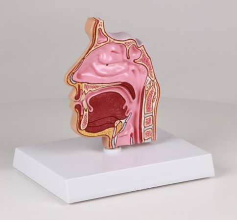
This cut-away model depicts a near median section through the nose and nasal passages. Details include nasal cavity, soft & hard palate, uvula and pharangeal tonsil. Back side shows ethmoid and maxillary sinus ...
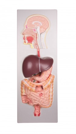
This life size model shows the human digestive tract from mouth cavity to rectum. The oral cavity, the pharynx and the first part of the esophagus are dissected along the medial sagittal plane. The liver is shown ...
