Quality Certyficate
street: Kolejowa 2, 30-805 Cracow


Home
3D anatomy models
Anatomical Models
Custom tools for patient education
Veterinary simulators
Anatomical Charts
Anatomical Table 3D
Medical simulators
Medical Equipment
Type:
 See our profile on Facebook
 Check our profile on Instagram
 Download a PDF file
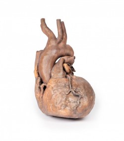
This 3D model represents a ‘normal’ sized adult heart with light dissection to the epicardium to expose the coronary arteries and cardiac veins.
At the base of the heart, the terminal part of the superior ...
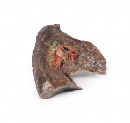
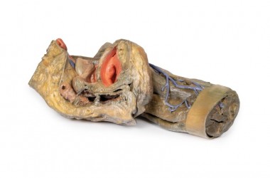
The hilum of a lung is the point at which visceral and parietal pleura meet and functions with the pulmonary ligament as the lungs only connection with the rest of the body. This connection includes the Pulmonary ...
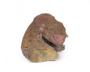
The lung has been dissected following a parasagittal plane, removing the mediastinal surface. Ordinarily, the pulmonary arteries, veins and bronchi can be observed entering the lung in the hilum – but the primary ...
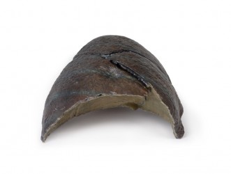
This 3D model represents the complimentary section to the TW 63 right lung hilum 3D model within our series and provide a direct contrast to the TW 61 left lung section. While expressing few discrete features, this 3D ...
The hilum of a lung is the point at which visceral and parietal pleura meet and functions with the pulmonary ligament as the lungs only connection with the rest of the body. This connection includes the Pulmonary ...
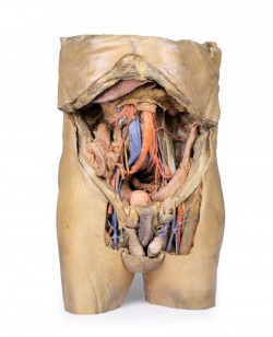
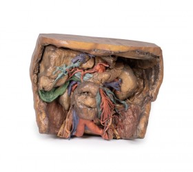
This 3D model represents one of the largest and most complex in the series, consisting of a partial torso from the diaphragm to the proximal thigh with a complete abdominal cavity preserving varying levels of ...
Clinical History
The patient was a 51-year old woman who had a cerebrovascular accident resulting in a left hemiplegia 2 years prior to death. At necropsy, she had severe generalized atherosclerosis and an old ...
Clinical History
A 29-year old male presented with a 22 month history of headaches and blurred vision. Examination revealed a bi-temporal hemianopia and a left VIth nerve palsy. Skull X-ray showed erosion of most of ...
Clinical History
A 56-year old male underwent a total gastrectomy and splenectomy for gastric adenocarcinoma. Over a period of two months he developed a progressively unsteady gait, increasing weakness of his left hand ...
Clinical History
A 50-year-old alcoholic was admitted with a 2-week history of weakness and shortness of breath. At the onset of the illness he reported a productive cough, chest pain and blood-stained sputum. ...
Clinical History
A 22-year-old male presented with a 2-week history of generalised malaise, weight loss and bruised skin without any trauma. He recently developed 5 days of productive cough and fevers. He was ...
Clinical History
This patient died at the age of 58 years from post-operative complications following transurethral resection of the prostate. At the ages of 28 and 35, he had suffered two episodes of transient ...
Clinical History
A 56-year-old woman with 6 months of intermittent headache and vomiting was admitted to hospital comatose after a grand mal seizure, and failed to regain ...
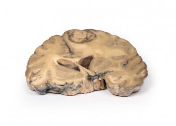
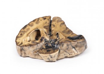
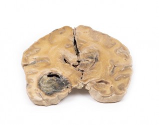
Clinical History
The patient was an 80-year old man who suddenly lost consciousness. On examination there was a right gaze palsy, a left hemiplegia and right hemiparesis.
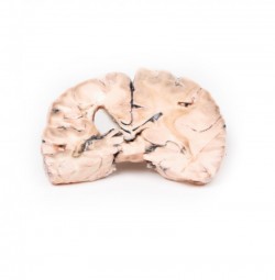
Pathology
The specimens are coronal ...
Clinical History
Five days before admission this 38-year old female experienced the sudden onset of pain behind the right eye, associated with a slow development of weakness of the left leg. Examination disclosed ...
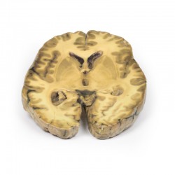
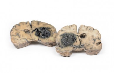
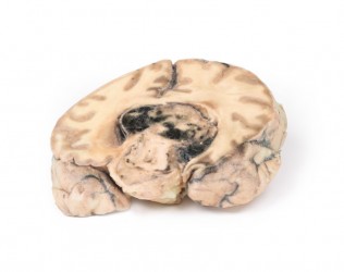
Clinical History
A 73-year-old female was admitted with new left-sided hemiplegia. On further questioning she revealed a 3-month history of headaches, nausea and deteriorating balance. CT brain revealed an ...
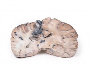
Clinical History
Over a 3-year period, a 57-year-old woman had intermittent frontal headache and memory disturbance with progression to psychiatric disturbance, and ultimately vomiting and meningeal signs. ...
Clinical History
A 56-year old male presented with a generalised seizure. He remained unconscious after this seizure and later died. A collateral history revealed 6 months of progressive confusion, short-term ...
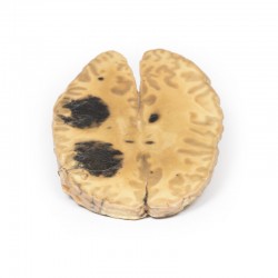
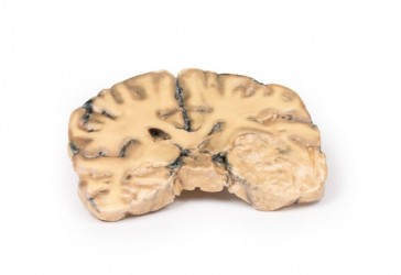
Clinical History
A 62-year-old woman presented with disorientation to time, place and person. Physical examination revealed no localised neurological signs. Radiological investigations revealed a space occupying ...
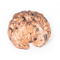
Clinical History
In the 1970s, a 31-year-old woman presented with severe headache and diplopia on a background of having a pigmented skin lesion (diagnosed as an invasive skin melanoma) removed from her neck 8 ...
Coeliac Trunk: Supplying the embryological foregut, the celiac trunk arises from T12 spinal level. Branches that can be observed in this specimen include the Left gastric artery arising from the left portion of the ...
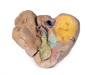
At the splenic hilum, the splenic artery and vein can be seen entering the spleen to supply and drain the organ. The opening of the splenic vein has been kept patent by the insertion of silicon tubing in the model. This ...
Diaphragm and Xyphoid Process: The diaphragm has been secured to the superior border of the dissected specimen with sutures to ensure an unobstructed view of the abdomen. The xyphoid process is in the middle of this ...
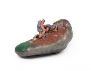
This 3D model is an isolated stomach with two dissection windows to expose the rugae and pylorus. A small portion of the terminal oesophagus is preserved at the cardiac region, and a small portion of the proximal ...
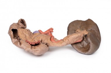
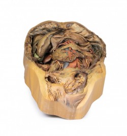
This 3D model preserves the deep foregut organs: the descending, horizontal and ascending duodenum, the pancreas, and the spleen. A small window in the duodenum has been opened to allow for a view of the plicae ...
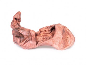
The size and shape of this specimen varies somewhat from a typical liver. It is less wedge-shaped and longer in the superoinferior dimension (on the posterior view this would translate to a greater vertical height). ...
This 3D model captures the internal surface of the anterior abdominal wall, a region oftentimes removed or damaged during dissection (and complimenting our A8 abdominal specimen where the anterior wall has been ...
This 3D model preserves a left pelvis divided at the midsagittal plane, and the proximal thigh to approximately the midthigh.
In the midsagittal section, the urinary bladder, uterus and vagina, and rectum can ...
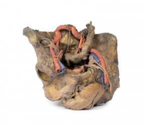
This 3D model presents a deep dissection and isolation of the pelvis from surrounding regions, particularly demonstrating visceral and neurovascular structures relative to deep ligaments and osseous ...
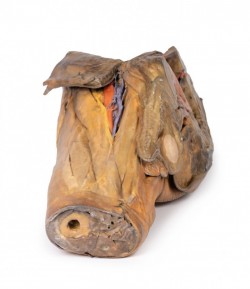
This 3D model preserves a right male pelvis sectioned just superior to the L5 vertebra and sectioned at the midsagittal plane, with the thigh preserved to near the midshaft of the femur. This specimen compliments our LW ...
Clinical History (pre access to CT and MRI imaging)
This 51-year old woman had surgery for breast carcinoma 2 years before presentation. Her main complaint was left-sided ataxia for the 2 weeks prior, and this had ...
Clinical History
This 70-year-old man with a past history of mild gastro-oesophageal reflux presented to the Alfred Hospital with a sudden onset of severe upper abdominal pain, which radiated to the left shoulder tip. ...
