Quality Certyficate
street: Kolejowa 2, 30-805 Cracow


Home
3D anatomy models
Anatomical Models
Custom tools for patient education
Veterinary simulators
Anatomical Charts
Anatomical Table 3D
Medical simulators
Medical Equipment
Type:
 See our profile on Facebook
 Check our profile on Instagram
 Download a PDF file
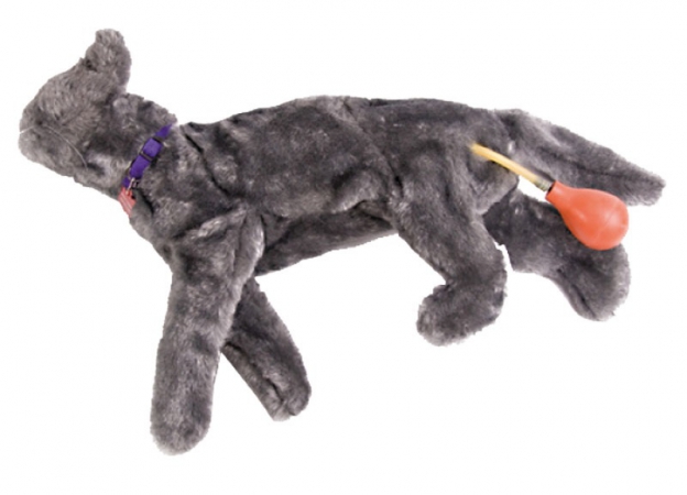
The manikin has realistic working lungs and an artificial pulse. Students learn to perform feline compressions and mouth-to-snout resuscitation. It is also designed to splint and allow bandaging. Parts can be cleaned ...
Description:
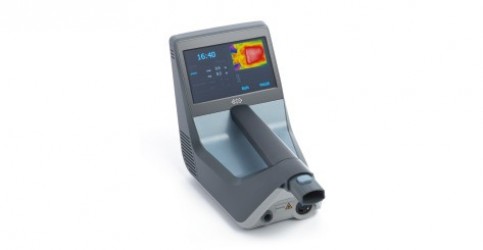
• 7” color touch screen
• Built-in thermal imaging camera
• Temperature sensor
• Distance sensor
• Thermal perception scan
• 6-joint arm for ease of use
• Precise application on large ...
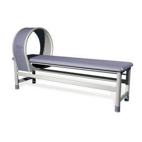
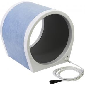
70cm, for BTL-4000/5000 Magnet
Magnetotherapy couch with sliding solenoid
Comfortable application of magnet therapy for both the patient and therapist
Main features:
Magnetotherapy couch with ...
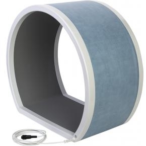
for BTL-4000/5000 Magnet ...
for BTL-4000/5000 Magnet
...
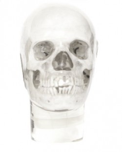
Human skull designed for radiographic examinations, embedded in a special, transparent material that does not cause image artifacts.
Main features:
The jaws are slightly open to allow dental panoramic ...

Clinical History
This 47 year old woman was admitted with terminal carcinomatosis. On examination a hard liver and a right pelvic mass were palpable. She had been ill with constitutional symptoms for months and she ...
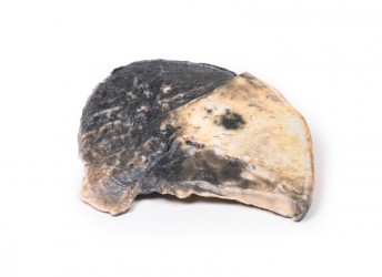
Pathology
The specimen is a parasagittal section of the right lung and the boundaries between the three lobes are visible. The entire upper and middle lobes are congested and hyperaemic* causing the darker appearance. ...
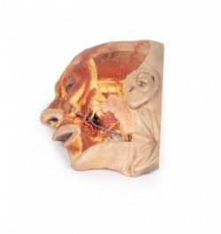
This 3D model presents a superficial dissection of a left face anterior to the ear with false colouring highlighting a series of neurovascular structures alongside the superficial muscles of facial expression. This ...
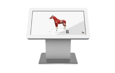
Digital, mobile platform on road wheels for teaching animal anatomy. The veterinary anatomical table is an innovative, interactive and holistic approach to veterinary education. Equipped with the world's most popular ...
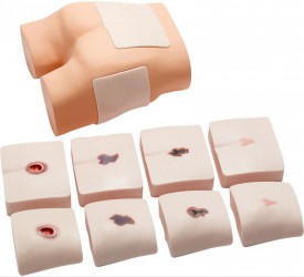
This unique training phantoms allows training of ultrasonic detection of DTI (deep tissue injury). The phantom has two replaceable pads in two locations where DTI typically occurs easily. One pad represents the ...
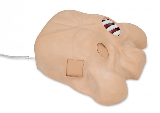
Now there is a manikin designed specifically to teach the theory, anatomy, and skills needed to manage pre-hospital chest trauma, as well as ongoing chest tube maintenance. The right side of the manikin has two ...
This unique model combines the most important procedures in thorax trauma treatment. It is made of life-like silicon material and provides realistic training feeling to the student.
The following procedures can be ...
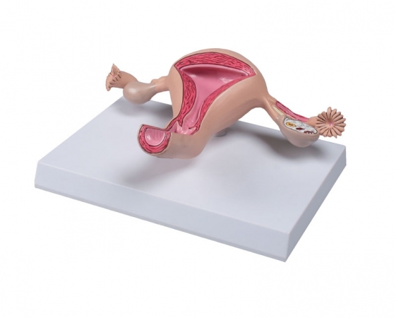
Life size model of a healthy uterus. Anatomy is shown in detail, structures are carefully painted by hand. Cervix, endocervical canal, uterine cavity are sectioned to show endometrium and myomterium. Also one ...
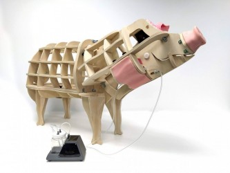
It is one of the modules for the overall pig model (MZ02661). It is used to teach how to draw blood from a pig's neck. It is intended for students. This model is completely anatomically correct, and the materials from ...

This 3D model provides a view of the isolated brainstem anatomy from the midbrain to the medulla oblongata, and compliments the other diencephalon/brainstem 3D model (BR 10) in our series. Rostrally, the 3D model has ...
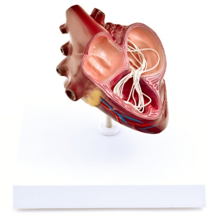
This anatomical model depicts an average-sized dog’s heart that is infected with Dirofilaria immitis. The cut-away model shows the structures of the heart where adult heartworms can usually be found in the event ...
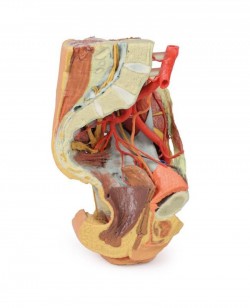
This 3D printed female left pelvis and proximal thigh preserves both superficial and deep structures of the true and false pelves, inguinal region, femoral triangle, and gluteal region. The specimen has been sectioned ...
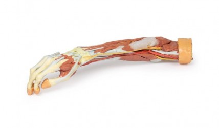
This 3D print of a superficially dissected right upper limb specimen displays a mixture of the vascular, nervous and muscular anatomy of the distal arm, forearm and hand. In the distal arm and elbow/cubital fossa ...

This 3D printed specimen compliments our dorsal dissection specimen (AM01273) by presenting a ventral deep dissection of axial anatomy from the head, neck, axillae, thorax, and abdomen to the proximal portion of the ...
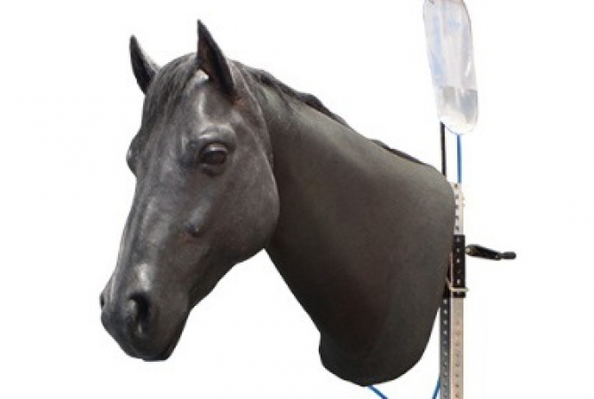
This advanced and realistic head simulator of equine on a tripod, designed for training and improving the skills of venipuncture and intramuscular injections.
Main features:
Jugular venipuncture with palpable ...
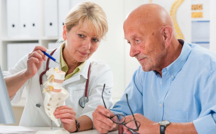
Patient's education increases the effectiveness of therapy!
Patients wants to know more about the disease they are suffering from. Also they want to know the way of therapy. Help your customer to improve the patient ...
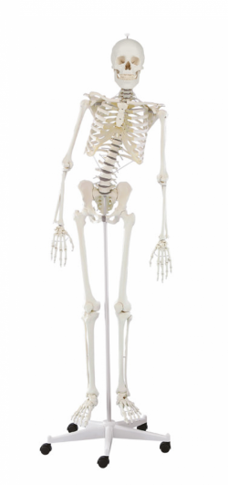
This therapy skeleton with a moveable vertebral column is ideal for anyone who not only wants to learn anatomy, but also wishes as a therapist to understand or explain the connections between movements, postures and ...
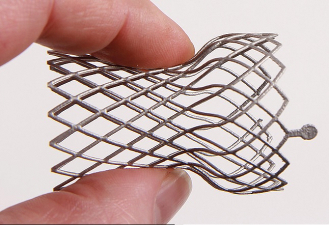
Medical Device Replica
We offer a cost-effective alternative to your original device. The replicated devices can be used for marketing purposes or patient education Service.
Services
Custom anatomical ...
This model shows the wrist and carpal tunnel.
The following ligaments are shown, among others:
Retinaculum flexorum, Lig. pisometacarpum, Lig. pisohamatum, Tendo m. flexoris carpi radialis, Lig. ulnocarpale palmare, ...
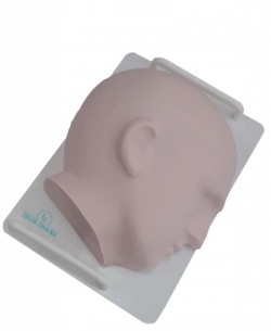
The Facial simulator contains the following muscles:
Frontalis, Temporalis, O/Oculi, Risorius, Zygomaticus major & minor, Levator Labii, Buccinator, Alaeque Nasi, Orbicularis Oris, Labii Inferioris, Mentalis, ...
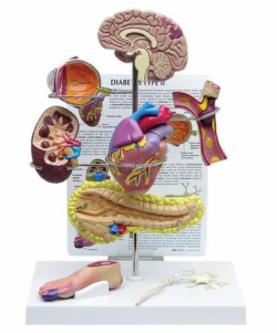
The includes miniature brain, eye, heart, kidney, artery, pancreas, neuron, and foot models. Education card illustrates effects associated with Type II Diabetes: stroke, ocular pathology, hypertensive heart disease, ...

The model simulates the correct and incorrect way of lifting weights for the spine. Ideally suited for demonstration purposes in cabinet conditions.
Size: 23 x 15 x 15 cm ...
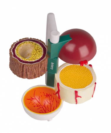
Four piece model indicating structures and organs with vascular effects due to diabetes.
Includes sectioned model of Bowman‘s capsule (kidney), artery, nerve, eye (posterior section).
Full model size without ...
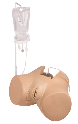
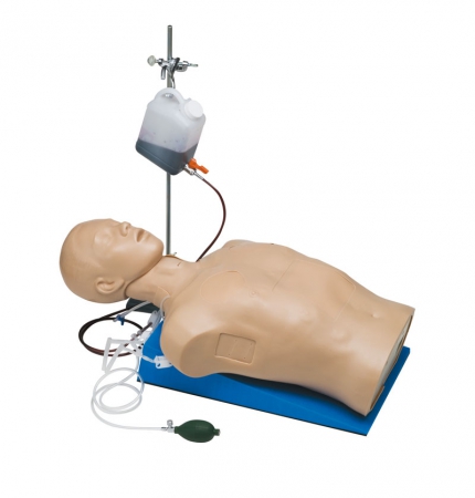
A professional training simulator for learning catheterization. This model is perfect for patient education.
Main features:
The innovative bladder is designed to provide a normal, anatomical and ...
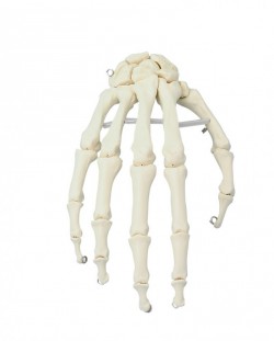
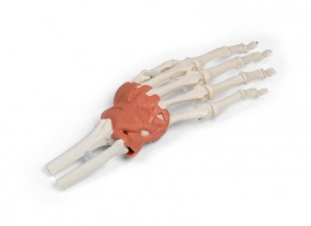
A life-size anatomical model showing the skeletal structure of the hand. It Shows all palm bones which are individually mobile-mounted on wire
It is possible to perform translational movements in individual hand ...
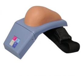
This high-quality I.M. simulator represents a right upper arm with all important anatomical palpable landmarks such as acromion and humerus. The realistic anatomy allows for placing correct intramuscular ...
