Quality Certyficate
street: Kolejowa 2, 30-805 Cracow


Home
3D anatomy models
Anatomical Models
Custom tools for patient education
Veterinary simulators
Anatomical Charts
Anatomical Table 3D
Medical simulators
Medical Equipment
Type:
 See our profile on Facebook
 Check our profile on Instagram
 Download a PDF file
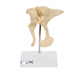
The three smallest bones that are joined to each other in the human body are located in the middle ear and are referred to as the auditory ossicles: malleus (hammer), incus (anvil) und stapes (stirrup). Their job is to ...
Contains:
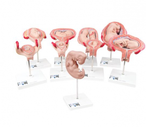
• Embryo approx. 4 weeks old, 25 times life-size
• 1st Month Embryo,
• 2nd Month Embryo,
• 3rd Month Embryo,
• 4th Month Foetus (Transverse Lie)
• 5th Month Foetus (Breech Position)
• 5th ...

This one-piece model, 150 times life size, is an important tool to study the histology of the most important organ of the digestive system: the stomach. All the different layers from the epithelium to the serous coat ...
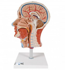
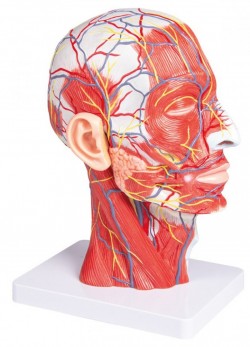
This high quality model represents the outer, superficial and the internal (median section) structures of head and neck. The half head with musculature is delivered on removable stand for easy display in a classroom or ...
The synapse model features the endoplasmic reticulum, mitochondria and the membranes of the synaptic gap. Also depicted by the synapses model are 5 smaller relief models of the main synapse variations. This synapse ...
Product information: Female genital model
The model represents the internal and external genitalia of a woman and is divided into four parts.
specifications:
dimensions: 14cm x 15cm x 12cm
displays 40 ...
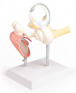
Enlarged approx. 15 times. The model shows instructive the organs of the middle and inner ear. The bony and membranous labyrinths are shown and the cochlea can be opened. On stand. ...
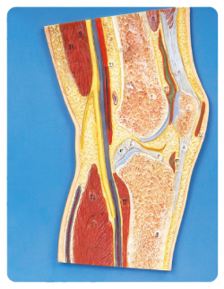
Product information: Cross section of the knee joint in a horizontal plane
A life-size model of the knee joint are visible, among others:
femur
quadriceps tendon
Throttle
patella
cavity of ...
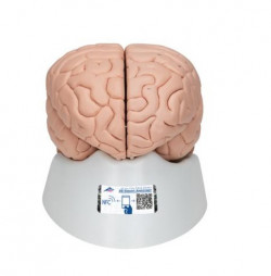
This 4-part brain is medially divided. All structures of the brain are hand-painted, numbered and identified in a product manual.
The brain's right half can be disassembled into:
Frontal with parietal lobes
...

This medially divided deluxe brain model shows the brain arteries. The basilar artery of the brain is removable for added detail.
Both halves of this high quality brain model can be disassembled into:
...
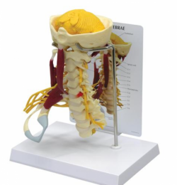
The Deluxe muscled Cervical Spine is a full size model features brain stem, occipital bone, atlas and axis through C7, with herniated disc, T1 and T3. This model has a soft cerebellum and full nerve with right side ...
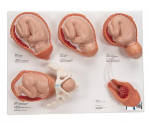
5 stages of human birth, mounted individually on bases:
Fetus in womb, cervix closed
Fetus in womb, cervix open
Fetus in womb, start of head passage
Fetus in womb and pelvis, finish of head ...
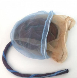
A great teaching tool, this model of the placenta, cord, amnion, and chorion is perfect for childbirth education courses.
An educator essential, this model is perfect for use on its own or in combination with ...
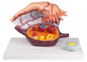
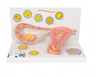
This 2part, 5times life size model shows the anatomy of the human fallopian tube and ovary. The follicles are shown at different stages of maturation, from the primary follicle to the corpus albicans. A primary ...
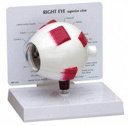
Oversized normal eye model with split shell construction to allow for viewing inner anatomy including optic nerve, disc, macula, retina, central retinal artery and vein. Lens and cornea are removable.
Model size: 5 x 3 ...
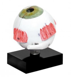
Model of the human eye, enlarged to a scale of 3 to 1, including the cornea, the lens and glass bodies for demonstrating the fundamentals of ultrasonic biometry. The biometric ratios in the human eye (distance between ...
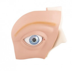
This eye model shows the eyeball with optic nerve in its natural position in the bony orbit (floor and medial wall). Additionally, this eye model shows the relation between eye, bones, muscles, and outer structures of ...
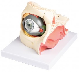
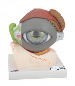
This 13-part model, enlarged approximately 2,5x life-size, shows the anatomy of the human eye.
The orbit can be opened into 3 parts to reveal the internal structures:
eyelid with lacrimal gland and duct
...
This anatomical model is great for studying the anatomy of the human eye. Eye on base of bony orbit.
Removable parts of the anatomical human eye model include:
Upper half of the sclera with cornea and eye muscle ...
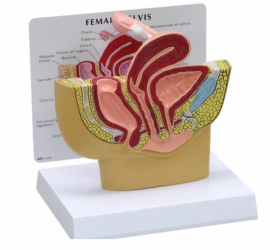
Reduced size, mid-sagittal cross-section of the pelvis showing female anatomy, including the ovary and fallopian tube.
Model size: 6.25 x 2.75 x 5.5"
Base: 6.5 x 5" ...
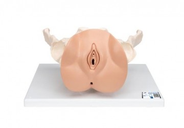
This female pelvis skeleton model is especially suitable for studying female genital organs in the context of their anatomical position in the the pelvis. It consists of female pelvis with a movable symphysis, hip bone, ...
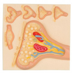
The model illustrates schematically how the ovum matures, how ovulation and fertilization occur and how the fertilized ovum develops to the stage where it embeds itself in the womb wall to begin the growth into an ...
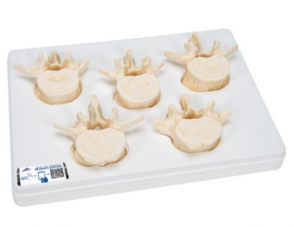
A worldwide unique high quality anatomical replica of original lumbar vertebra bones, which represents the finest anatomical structures in detail.
Manufactured in excellent quality from 3B BONElike™ material, which ...
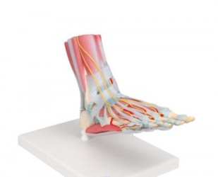
This anatomically detailed model of the foot and lower leg can be disassembled into 6 removable parts for detailed study of the foot and ankle. The foot skeleton features not only the bones but also the muscles, ...
Life size model shows the right half of the human head and neck, sectioned along the sagittal plane. A superficial dissection exposes the facial muscles, the superficial blood vessels and nerve branches of the face ...
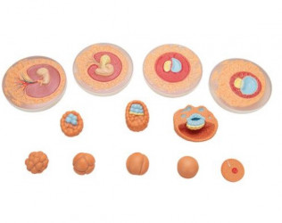
The model represents the development of the human germ cells from fertilisation until the end of the 2nd month of pregnancy in 12 stages. Each stage can be removed from the common stand as an individual part and can be ...
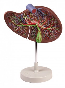
Enlarged 1.5 times, this model shows a liver that is dissected to expose the internal distribution of arteries and veins, the portal vein and the bile duct. Mounted on stand.
Size: 15 x 26 x 12 cm, Weight: approx. 1 ...
This 3D model is a midsagittal hemisection through a whole brain, preserving the right side anatomy and deep brain structures and spaces visible in the midline. In lateral view, the right cerebral and cerebellar ...
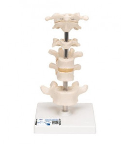
Consisting of atlas, axis, another cervical vertebra, two thoracic vertebrae with inter-vertebral discs and one lumbar vertebra. On stand, removable.
Every original 3B Scientific® Anatomy Model gives you direct access ...
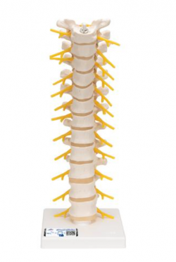
This spinal replica consists of the 12 thoracic vertebrae with intervertebral discs, thoracic nerves and spinal cord. This quality thoracic spinal column is affordable and anatomically correct. Spinal column delivered ...
This spinal cord model shows a segment of the upper thoracic spinal cord and is laterally and longitudinally divided showing spinal nerve roots. It is approximately 6 times life size and comes on a baseboard. ...
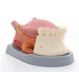
This tongue model shows the lower jaw up to the second molar, the tongue with mouth floor musculature in median section and the right sublingual and submandibular gland.
Tongue on removable base.
Every original 3B ...
