Quality Certyficate
street: Kolejowa 2, 30-805 Cracow


Home / 3D anatomy models / 3D lower limb
3D anatomy models
Anatomical Models
Custom tools for patient education
Veterinary simulators
Anatomical Charts
Anatomical Table 3D
Medical simulators
Medical Equipment
Type:
 See our profile on Facebook
 Check our profile on Instagram
 Download a PDF file
3D anatomy models / 3D lower limb
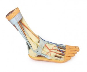
This 3D print records the anatomy of a right distal leg and the deep structures of the plantar surface of the foot. Proximally, the tibia, fibula, interosseous membrane, and leg muscles are discernable in ...
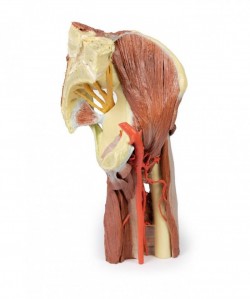
This 3D printed specimen presents a deep dissection of a left pelvis and thigh to show the course of the femoral artery and sciatic nerve from their proximal origins to the midshaft of the femur. Proximally, the pelvis ...
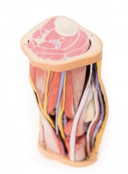
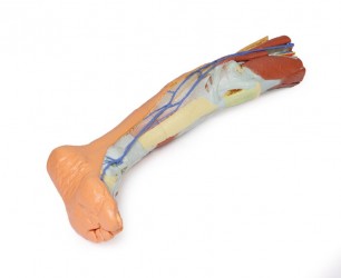
This 3D printed specimen preserves the distal thigh and proximal leg, dissected posteriorly to demonstrate the contents of the popliteal fossa and surrounding region. The proximal cross-section demonstrates the ...

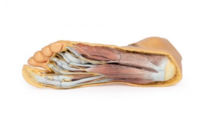
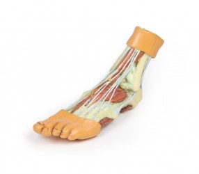
This 3D printed specimen provides a view of deep plantar structures of a right foot. Medially, the cut edge of the great saphenous vein is visible within the superficial fascia, just anterior to the cut edges of the ...
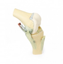
This 3D printed specimen demonstrates the ligaments of the knee joint with the leg in flexion. In the anterior view, with the patella and part of the patellar ligament removed, the medial and lateral menisci and ...
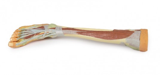
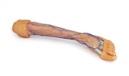
This 3D printed specimen presents both superficial and deep structures of a right distal leg and foot. Proximally, the posterior compartment of the leg has been dissected to remove the triceps surae muscles and ...
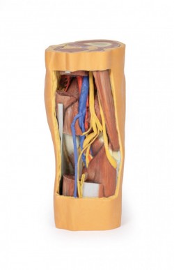
This 3D printed specimen preserves the distal thigh and proximal leg, dissected posteriorly to demonstrate the contents of the popliteal fossa and surrounding region. The proximal cross-section demonstrates the ...
This 3D printed specimen consists of a right partial lower limb sectioned just proximal to the knee joint and complete through a partially dissected foot exposing the structures on the dorsum. In the proximal cross ...
Lower Limb:
sectioned proximally near midthigh and continuous to the partially dissected foot. The transverse section through the thigh exposes the neurovascular structures of the anterior, medial and posterior ...
This 3D printed specimen presents a superficial dissection of a left lower limb, from just proximal to the knee joint to a complete foot. The skin and superficial fascia have been removed to display the superficial ...
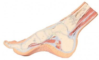
This 3D printed specimen provides a parasagittal cross-section through the medial aspect of the right distal tibia and foot, displaying the skeletal structures of the medial longitudinal arch of the foot and ...
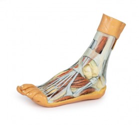
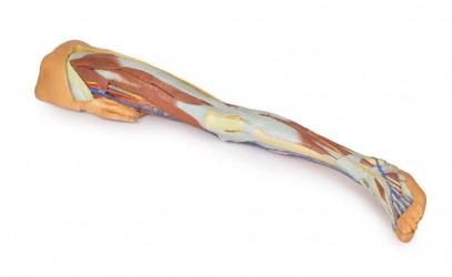
This 3D printed specimens preserves a mixed superficial and deep dissection of a left distal leg and foot. Posteriorly, the compartment muscles and neurovascular structures have been removed to isolate the ...
This 3D print records the anatomy of a right distal leg and the deep structures of the plantar surface of the foot. Proximally, the tibia, fibula, interosseous membrane, and leg muscles are discernable in cross-section. ...
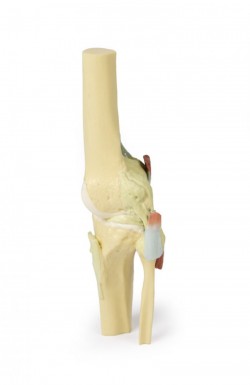
This 3D printed specimen demonstrates the ligaments of the knee joint with the leg in extension; it represents the same specimen as AM01281 knee joint printed in a flexed position. In the anterior view, with the patella ...
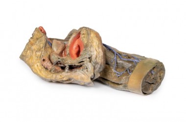
This 3D printed specimen preserves a superficial dissection of the lower limb musculature from the mid-thigh to mid-leg, as well as nerves and vessels of the popliteal fossa. The insertions of the muscles of the ...
This 3D printed specimen represents the remainder of the lower limb portions of our male abdominopelvic and proximal thigh specimen (MA01106)sectioned proximally near midthigh and continuous to the partially ...
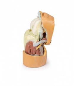
This 3D model preserves a left pelvis divided at the midsagittal plane, and the proximal thigh to approximately the midthigh.
In the midsagittal section, the urinary bladder, uterus and vagina, and rectum can ...

This 3D model preserves a right male pelvis sectioned just superior to the L5 vertebra and sectioned at the midsagittal plane, with the thigh preserved to near the midshaft of the femur. This specimen compliments our LW ...
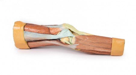
This 3D printed specimen displays a deep dissection of a left knee joint with the internal joint capsule structures relative to superficial tissues in a flexed position. The proximal cross-section through the distal ...
1
3D anatomy models - 3D lower limb
3D lower limb model as a teaching tool:
Anatomical models of the human lower limb are teaching aids presenting bones, muscles, blood and lymphatic vessels, and neural structures running in the lower limb region. Anatomically, the lower limb is divided into the hip girdle, thigh area, knee joint, lower leg area, ankle joint and foot. Taking into account the division, it should be noted that the human lower limb is a wealth of various anatomical structures and details, often running close to each other or crossing each other. From a teaching point of view, it is important to carefully visualize these structures in order to understand their functions as well as the mutual relations between them. During the teaching process, precisely made anatomical models, faithfully presenting the human body, work great. In this product category, we present anatomical models of the human lower limb created on the basis of CT radiological data and using modern 3D printing technology. OpenMedis also offers conventional anatomical models of the lower limb.
3D anatomical models of the lower limb - advantages:
It should be mentioned that the 3D anatomical models of the lower limb are made entirely of artificial material. This means that they do not wear out over time, creating the same, equal teaching conditions for all students. An additional advantage of 3D anatomical models of the lower limb is their very precise production, possible thanks to the use of radiological data from CT and 3D printing technology. These are high-fidelity anatomical models, which means that they accurately reflect the actual dissecting preparation on the basis of which they were made. Anatomical models of the lower limb present structures such as bones, muscles, nerves, blood and lymphatic vessels in their correct anatomical courses and positions. This is a great advantage and superiority of this type of anatomical models compared to conventional models that present the anatomy in a more exaggerated and illustrated way. Another important added value of 3D lower limb models is the fact that they do not require special storage conditions and can be used in regular seminar rooms. Additionally, it should be noted that these products do not require disposal and their service life is very long. There is no doubt that the colors of individual structures, tailored to the didactic purposes, facilitate learning anatomy by understanding the topography of individual structures and the mutual relationships between them.
3D anatomical models of the lower limb - application:
The presented 3D models of the human lower limb are an ideal educational aid for the process of teaching anatomy to students of medical faculties such as medicine, physiotherapy, nursing, and emergency medical services. Currently, it is a modern solution used at many universities around the world. 3D anatomical models of the human lower limb are perfect equipment for anatomical and biological laboratories and human anatomy museums. They are also perfect for courses and training in various fields of medicine. Anatomical models of the lower limb can also serve as a decorative element in teaching rooms in medical offices or constitute an element of thematic exhibitions on human anatomy.
Types of 3D lower limb anatomical models:
Our offer includes so-called anatomical models. conventional and unconventional human lower limbs. In this product category, we would like to interest you in 3D anatomical models of the lower limb, which faithfully present its anatomical structure as on real dissection preparations. There are anatomical models of the hip girdle, thigh models, lower leg models, foot models, and anatomical models of the entire lower limb.
- The anatomical model of the popliteal fossa (an important area in the teaching process) is very popular.
- Additionally, we recommend paying attention to the foot model, showing the dorsum of the foot and the muscles of the sole of the foot.
It is worth mentioning that our offer includes anatomical models presenting female and male anatomy. Anatomical models are made with the utmost care and from the best quality materials. Many of them are hand-finished. Thanks to the use of modern 3D printing technology, a unique line of 3D models was created. Based on radiological data, a series of models faithfully reflecting the human body were created. These advanced teaching aids are the equipment of anatomy laboratories of many Medical Universities in Poland. This is a modern solution used in many countries around the world.
How can we help?
OpenMedis is an experienced manufacturer and distributor of anatomical models on the Polish market. For many years, we have been successfully equipping anatomy laboratories of medical schools and universities, medical offices, schools and museums. If you need advice on equipment for the anatomy laboratory at your institution, we will be happy to advise you on the best product range to choose. Please contact us.
We also recommend 3D anatomical models of the upper limb and 3D torso models, which, combined with 3D models of the lower limb, will create a comprehensive set for learning the anatomy and physiology of the body.
