


Home / 3D anatomy models / 3D lower limb / Lower limb - superficial dissection with male left pelvis
Lower limb - superficial dissection with male left pelvis


Lower limb - superficial dissection with male left pelvis
 Download a PDF file Add to quotation - wish list
Product description: Lower limb - superficial dissection with male left pelvis

Lower Limb:
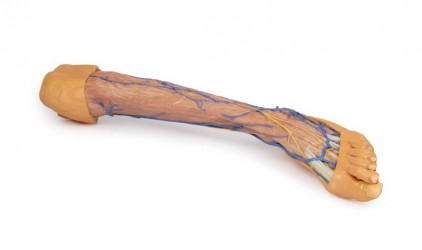
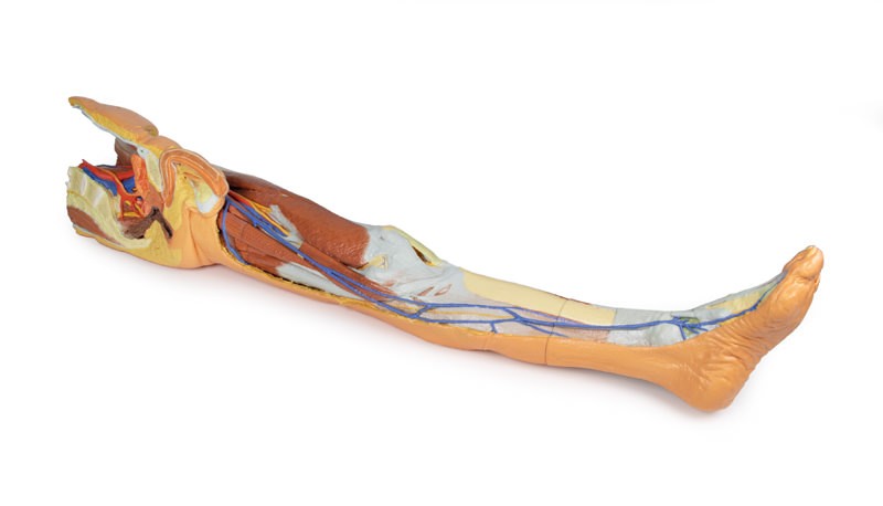
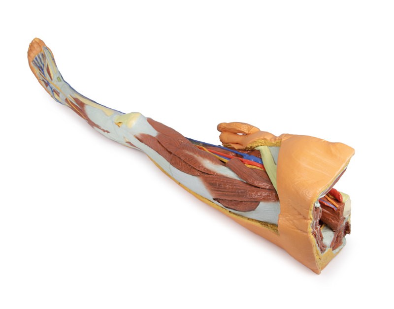
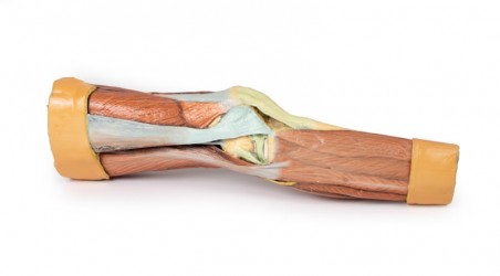
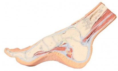
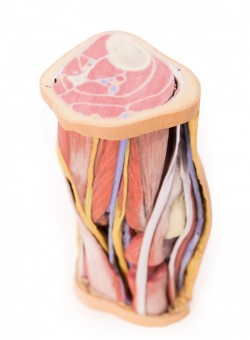
sectioned proximally near midthigh and continuous to the partially dissected foot. The transverse section through the thigh exposes the neurovascular structures of the anterior, medial and posterior compartments. This includes the great saphenous vein superficial to the terminal branches of the femoral nerve, femoral artery and vein in the anterior compartment, and perforating branches of the deep femoral artery in the medial and posterior compartments.
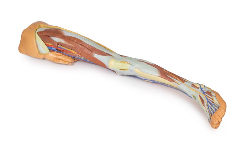
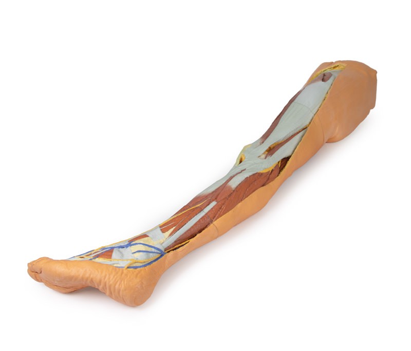
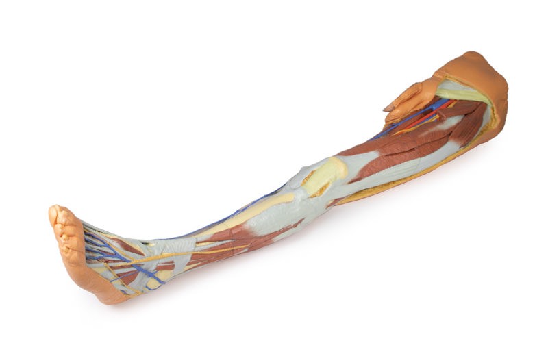
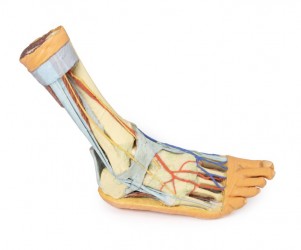
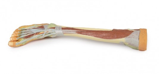
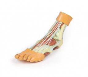
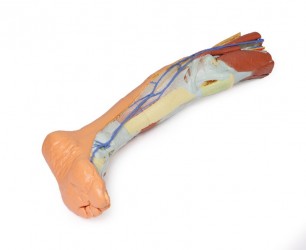
The remainder of the thigh, leg and dorsum of the foot have been dissected to demonstrate superficial structures and compartmental musculature, except the posterior aspect of the specimen which has been left undissected. The course of the great saphenous vein is displayed from the medial aspect of the thigh to the medial malleolus and the medial aspect of the dorsal venous plexus. The origin of the small saphenous vein from lateral branches of the dorsal venous plexus is also visible to the margin of the dissected superficial fascia near the lateral malleolus. The deeper femoral artery, vein and nerve branches are visible deep to the anterior compartment musculature (and a sectioned sartorius muscle) entering the adductor canal. Near the medial aspect of the knee joint the saphenous nerve is visible passing superficially near the great saphenous vein on the surface of the posterior crural fascia and terminating as the medial cutaneous nerve of the leg branches. On the lateral aspect of the leg the medial and intermediate dorsal cutaneous branches from the superficial fibular nerve are preserved passing onto the dorsum of the foot adjacent to the dorsal venous plexus tributaries.
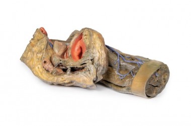
Male left pelvis:
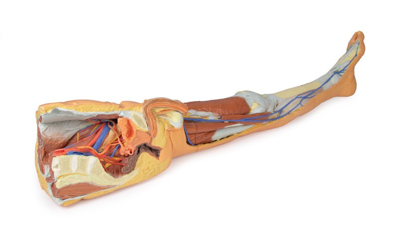
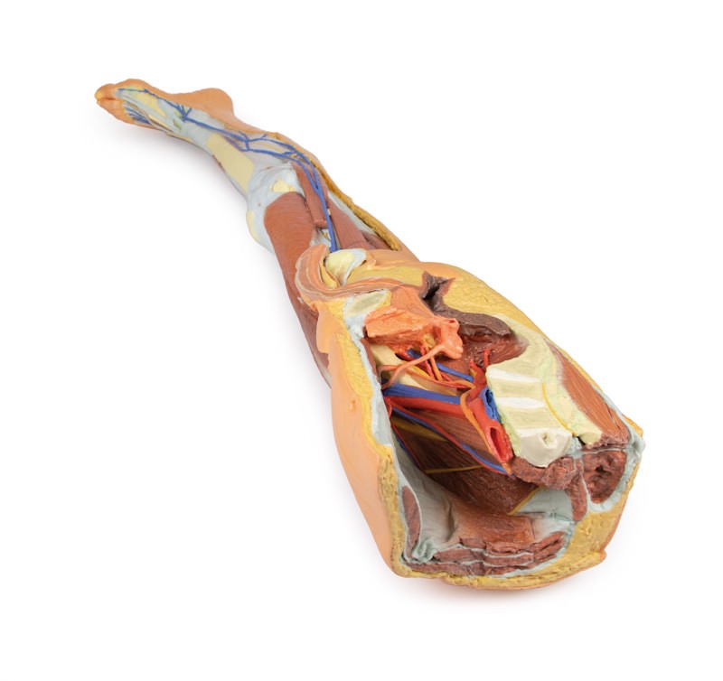
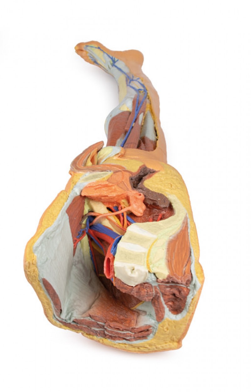
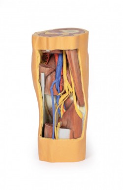
shows superficial and deep structures of the true and false pelves, inguinal and femoral region. In the transverse section, the epaxial musculature, abdominal wall musculature (rectus abdominis, external and internal abdominal obliques, transversus abdominis), psoas major and quadratus lumborum are visible and separated from each other and the superficial fat by fascial layers such as the rectus sheath and the thoracolumbar fascia. The psoas major muscle lies lateral to the external iliac artery, with the left testicular artery and vein lying on its superficial surface. More laterally (and moving inferiorly), the ilioinguinal nerve, the lateral cutaneous nerve of the thigh and the femoral nerve are positioned over the superficial surface of the iliacus muscle.
The left common iliac artery bifurcates at the level of the sacral promontory into the external and internal iliac arteries. This specimen does not possess a clearly defined anterior and posterior division of the internal iliac artery; instead, the terminal arteries sequentially radiate from the internal iliac. The lateral sacral, inferior rectal, inferior gluteal, internal pudendal, superior vesical, obturator and umbilical arteries (which terminates in the medial umbilical ligament) are visible adjacent to the sacral ventral rami. The inferior gluteal and internal pudendal arteries have not bifurcated in this view and track inferiorly over piriformis.
The deep circumflex iliac artery and vein can be seen passing deep posterior to the inguinal ligament, while the branches from the inferior epigastric artery and veins can be seen perforating rectus abdominis and the overlying rectus sheath. The left common iliac vein lies deep to the left common iliac artery; the obturator branch and the external iliac vein have been preserved.
In the midline the pubic symphysis and sagittal sections of the pelvic viscera are visible: from anterior to posterior, the bladder (receiving the left ureter, which passes over the iliac vessels at the level of the pelvic brim), the left seminal vesicles and vas deferens, and rectum (with surrounding external anal sphincter muscle). The pathway of the urethra is visible from the inferior pole of the bladder through the prostate gland, pelvic diaphragm and the corpus spongiosum of the penis. Inferior to the sectioned erectile bodies (corpus cavernosa and corpus spongiosum) lies the scrotum, where the skin has been removed to reveal the parietal tunica vaginalis.
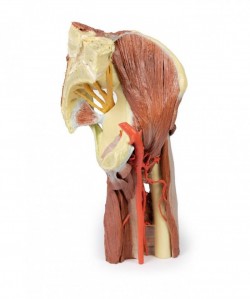
On the preserved proximal thigh the fascia lata has been removed to highlight the transition of the neurovasculature and musculature from the pelvic region. Superior to the inguinal ligament a window has been cut to reveal the underlying aponeurosis of the transversus abdominis muscle. From medial to lateral, the femoral vein and artery have been removed from the femoral sheath, and the termination of the femoral nerve lies superficial to the iliopsoas muscle.. The great saphenous vein can be seen coursing medially over the pectineus, adductor longus and gracilis muscles, while branches of the femoral nerve pass over the profunda femoris artery. The thigh musculature is visible, with the cut sartorius muscle overlying the iliacus muscles and the origins of anterior thigh muscles (rectus femoris, vastus lateralis, vastus intermedius, vastus medialis). The tensor fasciae latae can be seen inserting on the anterior border of the iliotibial tract, which extends over the lateral surface of the thigh. A window has been cut to expose the underlying gluteus medius muscle, which terminates at the lateral aspect of the greater trochanter.
Inquiry
Related products
