


Home / 3D anatomy models / 3D lower limb / Popliteal Fossa distal thigh and proximal leg
Popliteal Fossa distal thigh and proximal leg

Popliteal Fossa distal thigh and proximal leg
 Download a PDF file Add to quotation - wish list
Product description: Popliteal Fossa distal thigh and proximal leg

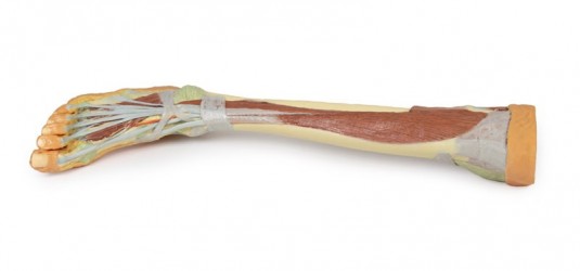
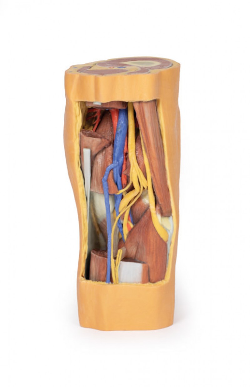
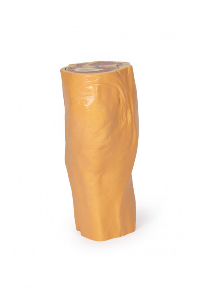
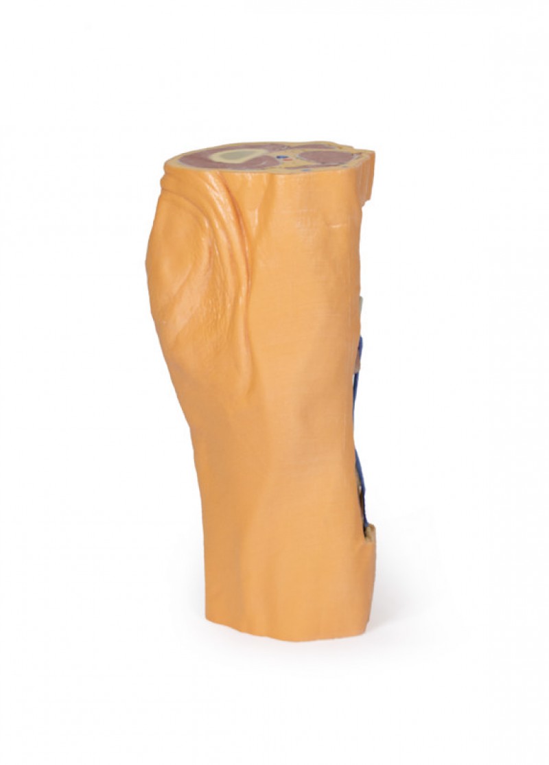
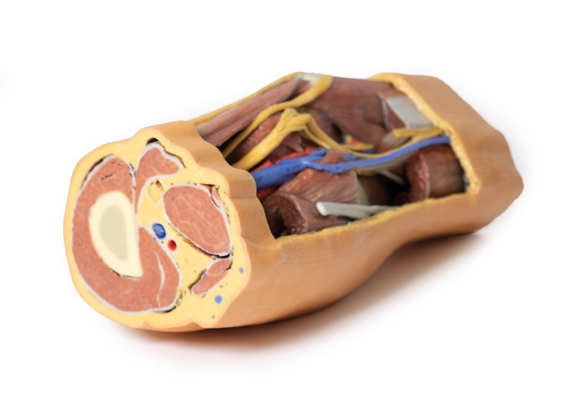
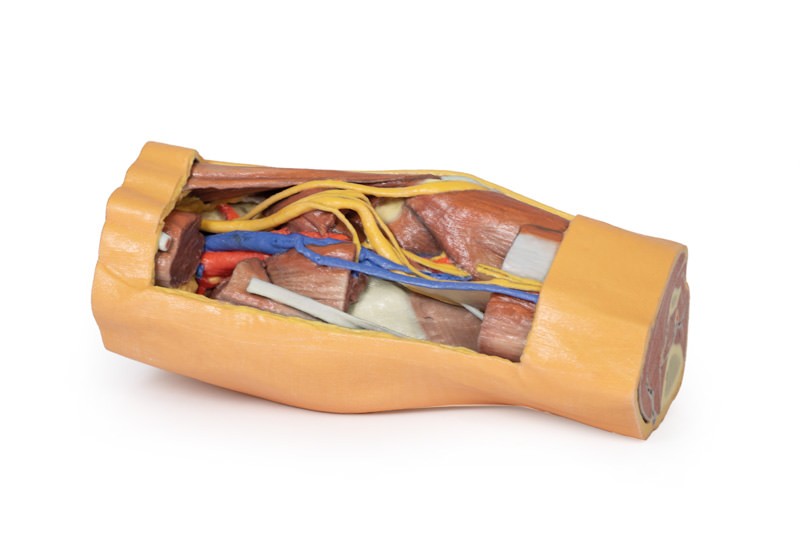
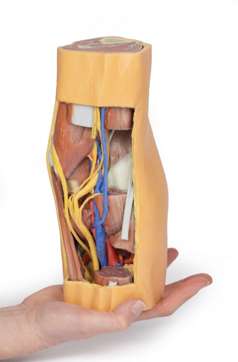
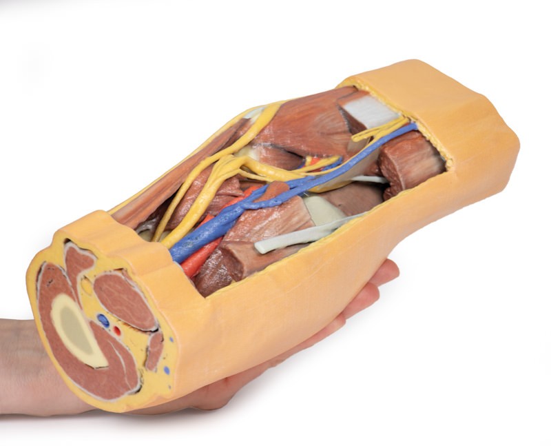
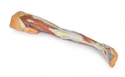
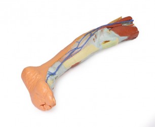
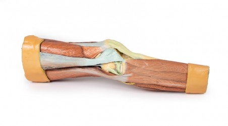
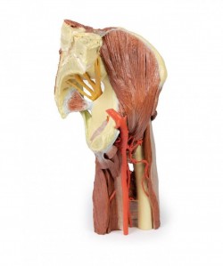
This 3D printed specimen preserves the distal thigh and proximal leg, dissected posteriorly to demonstrate the contents of the popliteal fossa and surrounding region. The proximal cross-section demonstrates the anterior, posterior and medial compartment muscles, with the origin of the popliteal artery and vein just as they have entered the popliteal fossa via the adductor hiatus. The sciatic nerve and great saphenous vein are also visible. The skin, superficial fascia, fascia lata and crural fascia has been removed posteriorly to demonstrate the course of the popliteal vessels, tibial nerve and common peroneal nerve. Medially, the semitendinosus and semimembranosus muscles have been sectioned to demonstrate the superior medial genicular artery and the medial head of the gastrocnemius. Distally, the medial gastrocnemius itself has been sectioned to expose the popliteus muscle and the tendon of the plantaris muscle. The course of the popliteal artery and vein can be traced through the fossa to the passage of the vessels deep to soleus. They are accompanied by the tibial nerve, with the lateral head of the gastrocnemius removed several muscular branches of the tibial nerve are visible in the fossa (as is the medial sural cutaneous nerve and the distal-most part of the lateral sural cutaneous nerve). Running in parallel, the common peroneal descends and passes laterally over the exposed soleus muscle to the neck of the fibula just distal to the attachment of the biceps femoris muscle. Deep to the biceps femoris, the superior lateral genicular branch can be observed passing towards the anterior compartment. The distal cross-section demonstrates the continuation of popliteal contents and branches. The great saphenous vein, small saphenous vein and sural nerves are visible within the superficial fascia. Between the muscles of the posterior, lateral, and anterior compartments are the neurovascular bundles of the leg (posterior tibial artery, veins and tibial nerve; peroneal artery and veins; anterior tibial artery, veins and deep peroneal nerve).
Inquiry
Related products
