


Home / 3D anatomy models / 3D lower limb / Lower limb - deep dissection
Lower limb - deep dissection

Lower limb - deep dissection
 Download a PDF file Add to quotation - wish list
Product description: Lower limb - deep dissection

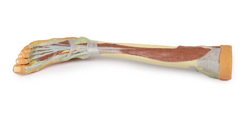
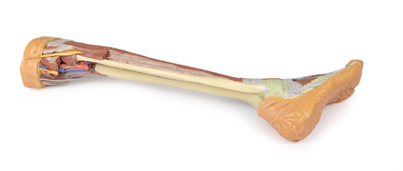
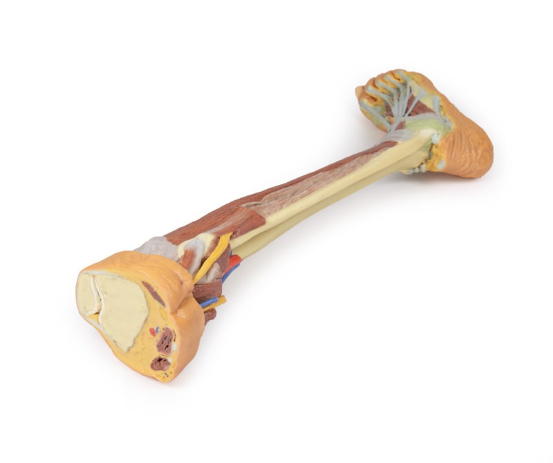
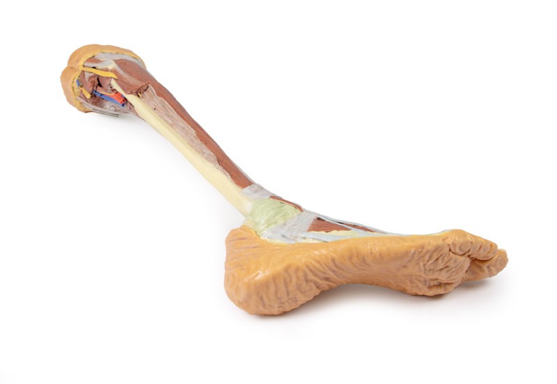
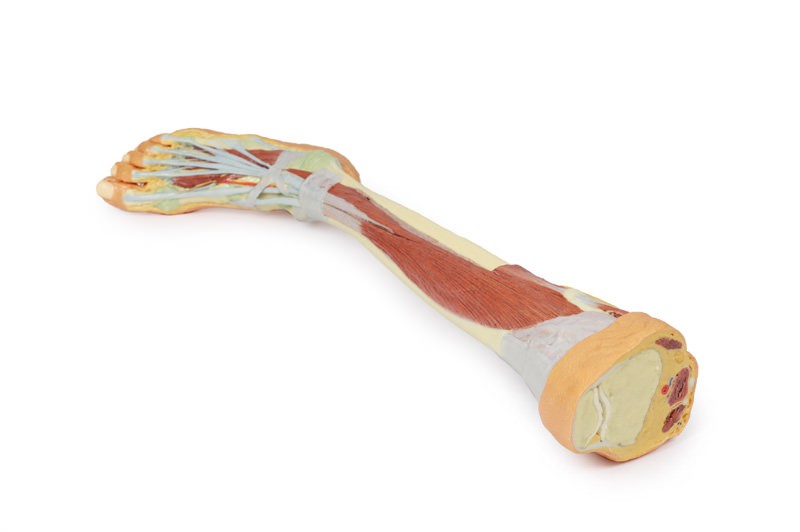
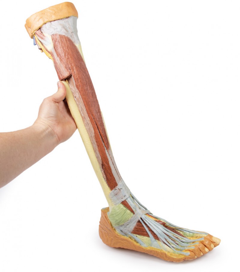
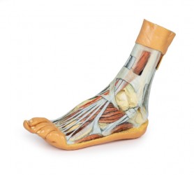
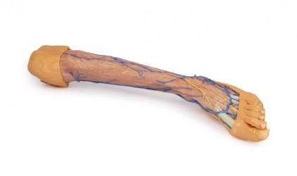
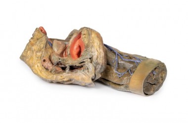
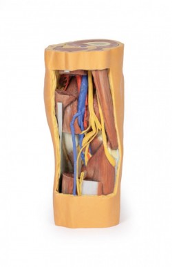
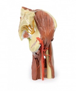
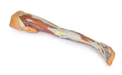
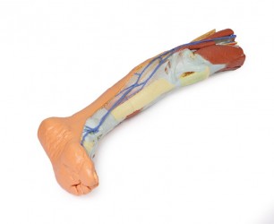
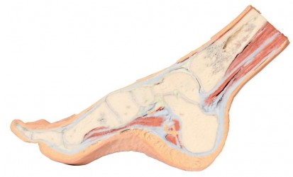
This 3D printed specimen consists of a right partial lower limb sectioned just proximal to the knee joint and complete through a partially dissected foot exposing the structures on the dorsum. In the proximal cross section, the patella articulates with the distal femur anteriorly, while the posterior portion of the specimen preserves structures within the superior portion of the popliteal fossa (including the popliteal artery, vein, and terminal portion of the sciatic nerve).
On the posterior aspect of the specimen distal to the knee joint, most of the musculature has been removed to demonstrate the passage of the neurovascular structures (common peroneal nerve, tibial nerve, posterior tibial artery, anterior tibial artery) relative to the deep musculature (e.g., popliteus muscle) and the interosseous membrane between the exposed posterior surfaces of the tibia and fibula. Medially the pes anserinus is visible inserting onto the medial aspect of the proximal tibia, while laterally the biceps femoris is seen inserting into the head of the fibula adjacent to the common peroneal nerve.
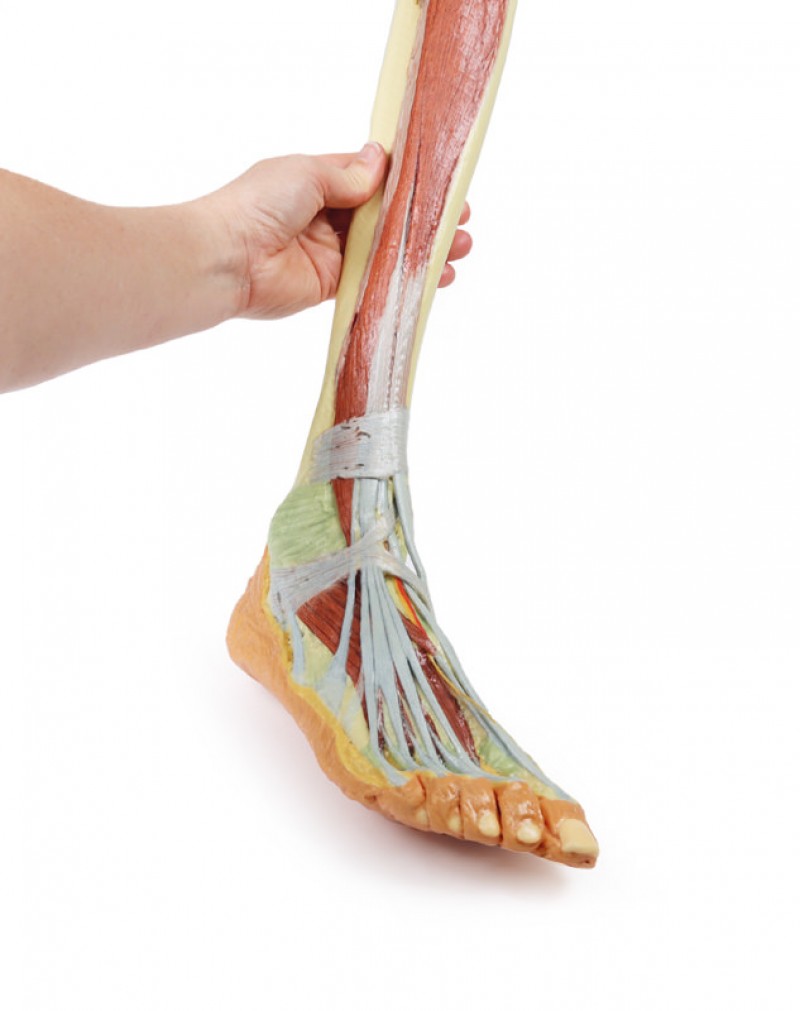
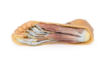
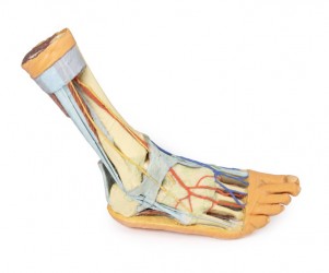
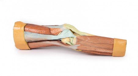
The bulk of the posterior compartment musculature (excepting the proximal deep posterior muscular origins and insertions and the distal tendons of the tibialis posterior, flexor digitorum longus and flexor hallucis longus) and the lateral compartment musculature (excepting the proximal portion of the fibularis longus muscle) have been removed to the ankle joint, while the anterior compartment musculature has been maintained and exposed deep to the crural fascia. Deep to the exposed posterior surface of the interosseous membrane the anterior tibial artery and vein can be seen passing distally through the anterior compartment. On the anterior and distal aspect of the specimen the tendons of the anterior musculature pass deep to the extensor and peroneal retinaculae and are visible passing to their respective insertions. The dorsalis pedis and the terminal portion of the deep peroneal nerve is visible lateral to the extensor hallucis longus tendon and medial to the extensor hallucis brevis tendons, and a well-developed extensor digitorum brevis is visible deep to the extensor digitorum longus and peroneus tertius tendons.
Inquiry
Related products