Quality Certyficate
street: Kolejowa 2, 30-805 Cracow


Home / 3D anatomy models / 3D anatomical models of pathologies and diseases
3D anatomy models
Anatomical Models
Custom tools for patient education
Veterinary simulators
Anatomical Charts
Anatomical Table 3D
Medical simulators
Medical Equipment
Type:
 See our profile on Facebook
 Check our profile on Instagram
 Download a PDF file
3D anatomy models / 3D anatomical models of pathologies and diseases

Clinical History
This 47 year old woman was admitted with terminal carcinomatosis. On examination a hard liver and a right pelvic mass were palpable. She had been ill with constitutional symptoms for months and she ...
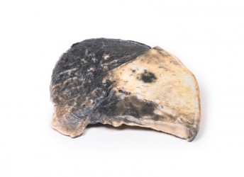
Pathology
The specimen is a parasagittal section of the right lung and the boundaries between the three lobes are visible. The entire upper and middle lobes are congested and hyperaemic* causing the darker appearance. ...
Clinical History
A 49-year old male presents with a 6-week history of malaise, urinary frequency and haematuria for 6 weeks. Further questioning revealed intermittent left flank pain. Abdominal ultrasound showed ...
Clinical History
A 54-year-old man presented to hospital with 12 hours of severe colicky pain, nausea and vomiting. On history, he was noted to have had a 3-year history of intermittent right subcostal pain for ...

Clinical History
A 5-year old male presents with a history of constipation since birth. A barium enema showed a constricted rectum with a dilated sigmoid colon. Surgical resection of constricted section of bowl ...
Clinical History
An 82-year old female presents with an episode of melena (dark tarry faeces). She had a 6-month history of dyspepsia and nausea. Recently she had noted weight loss and early satiety. Soon after ...
Clinical History
A 45-year old male presented with a lump in his left supraclavicular area. The swelling had been increasing in size over 6 months. Excision biopsy of the lump showed Hodgkin lymphoma (HL). Ten months ...
Clinical History
This 70-year-old man with a past history of mild gastro-oesophageal reflux presented to the Alfred Hospital with a sudden onset of severe upper abdominal pain, which radiated to the left shoulder tip. ...
Clinical History
This 56-year old female suffered from emphysema and gave a 2-year history of increasing shortness of breath on exertion associated with recurrent attacks of bronchitis. On examination, she had a BP ...
Clinical History
A 10-year-old girl with a known congenital heart was admitted for surgical repair because of the recent onset of cyanosis and cardiac failure. On examination, she was breathless with a blood pressure ...
Clinical History
A 64-year old woman presented with a story of chest pain for 5 months, associated with breathlessness and wheezing for 4 months. On examination, she was dyspnoeic, with an expiratory wheeze, left-sided ...
Clinical History
This male child had a cardiac murmur discovered at birth. He remained well until the age of 10 months. Two weeks before admission to hospital, he became languid, developed a slight cough, and suddenly ...
Clinical History
This 11-year-old female had an 18-month history of hydatid disease (see below). In total, 17 cysts were removed from the child’s brain at craniotomy on three occasions, and subsequently cysts were ...
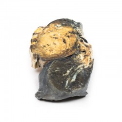
Clinical History
A thin 42-year-old American tourist was found dead in his hotel bedroom. A coroner’s autopsy was performed.
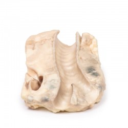
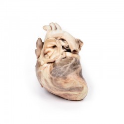
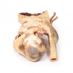
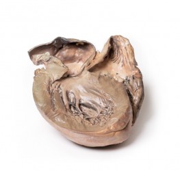
Pathology
This is a longitudinal section through the heart displaying the left and ...
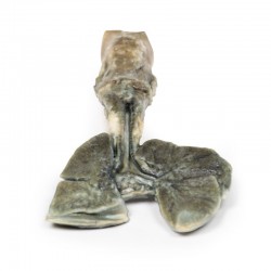
Clinical History
A 21-month old boy was admitted with a history of exhaustion and exertional dyspnoea for the previous 2 to 3 months. During this time there had been several attacks of acute dyspnoea each lasting up to ...

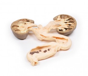
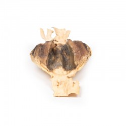
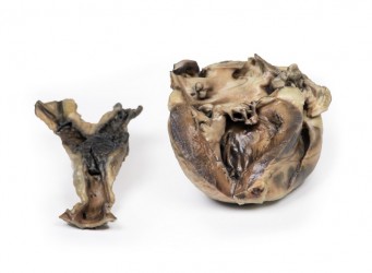
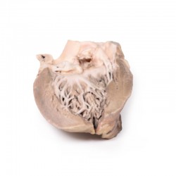
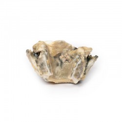
Pathology
The specimen is partial horizontal 1.5cm slice through the plane of the left atrium whose smooth internal lining together with the left auricular appendage and part of the left ventricle are visible on the ...

Clinical History
The patient was a 52-year old female with increasing dyspnoea. She gave a past history of fever with flitting joint pains in childhood following a sore throat. On examination: cyanotic, pulse showed ...
Clinical History
A woman who swallowed a chop bone during lunch collapsed later in the afternoon and suffered a massive haematemesis. At laparotomy, the stomach was filled with fresh blood but the cause was not ...
Clinical History
A 15-year old boy with cough and sputum developed a hectic (spiking) fever and chest pain a few days before being admitted in a comatosed condition. Examination revealed an early diastolic murmur at ...
Clinical History
A 61-year old male presents with exertional anginal chest pain and dyspnoea. He has had these symptoms for 6 years with increasing severity. On examination, he is cyanotic and tachycardic with a ...
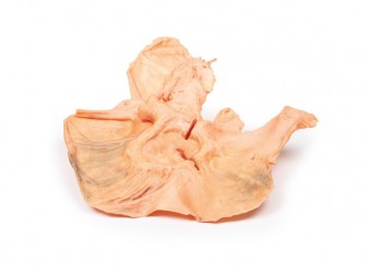
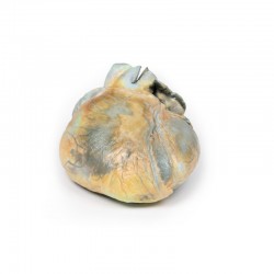
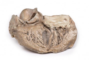
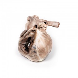
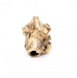
Pathology
The heart displays both ventricles from the posterior aspect. There is a prominent saccular dilatation of the thoracic ascending aorta, which shows several atherosclerotic plaques and posteriorly is seen to ...
Clinical History
A 47-year-old male presents with a 13-month history of dysphonia and odynophagia at the level of his thyroid cartilage. He has a significant smoking history. Investigations revealed a laryngeal tumour. ...
Clinical History
A 60-year old male presents with a 6-week history of globus (i.e. the feeling of a lump in his throat) and dysphonia. On examination, he had enlarged cervical lymph nodes. Investigations discovered a ...
Clinical History
A 74-year old male presented with a 2-months history of dysphagia, dysphonia and weight loss. He had a history of heavy alcohol consumption and smoked 40 cigarettes per day for 40 years. Investigation ...

Clinical History
A 89-year old male presents with an episode of large haemoptysis. He has a history of diabetes and immunosuppression secondary to steroid treatment for rheumatoid arthritis. Further history reveals a ...
Clinical History
A 37-year old male patient presents with a 1-month history of lethargy, cough and weight loss. He had a history of an orchiectomy 18 months previous for a testicular tumour. Then 12 months post-op he ...

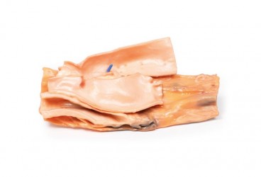
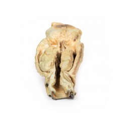
Pathology
The specimen is a parasagittal section of the left lung. There are patchy regions of focal consolidations and discolorations caused by congested and hyperaemic lung tissue distributed within both lobes; ...
Clinical History
A 32-year-old female G3P0 (gravida 3, para 0‘ – i.e., has had two pregnancies, with neither of the embyros surviving to a gestational age of 24 weeks) presents in preterm labour at 25 weeks ...

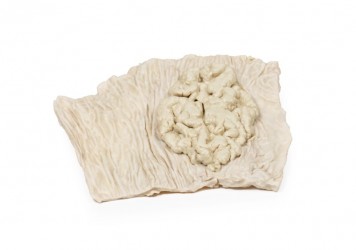
Clinical History
A 74-year old male presented with increasing shortness of breath and haemoptysis. Further history reveals 20kg weight loss in 6 months, night sweats and a chronic cough. He has recently moved from a ...
Pathology
The specimens from this case consists of two segments of sigmoid colon. The mucosa of the bowel is studded with numerous sessile and pedunculated partially pigmented polyps up to 1.5 cm in maximum diameter. ...
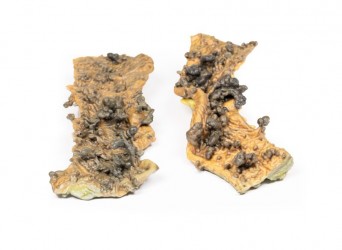
Clinical History
A 70-year old man was admitted for investigation of muscular weakness and the passage of large amounts of mucus per rectum. The patient was found to be hypokalaemic. A tumour of sigmoid colon was ...
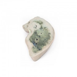
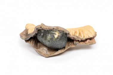
Pathology
A slice of liver reveals the characteristic yellow/grey and greasy appearance on one side. On the other side the appearance is restricted to the outer margin whilst the central area displays darker ...
3D anatomy models - 3D anatomical models of pathologies and diseases
3D anatomical models of diseases and pathologies - an excellent teaching tool:
3D anatomical models of diseases and pathologies are irreplaceable scientific aids that allow for better understanding and study of various aspects of diseases and pathologies in three dimensions. These advanced 3D models are based on real medical data and 3D printing technology, which enables faithful reproduction of disease lesions and anatomical anomalies. These models serve as an invaluable tool in both medical education and diagnosis and treatment. Our offer includes, among others: models of brain, heart, lung and kidney diseases.
3D anatomical models of diseases and pathologies - advantages:
It should be mentioned that 3D anatomical models of diseases and pathologies are made entirely of artificial material. This means that they do not wear out over time, creating the same, equal teaching conditions for all students. An additional advantage of 3D anatomical models of diseases and pathologies is their very precise execution, possible thanks to the use of radiological data from CT and 3D printing technology. These are high-fidelity anatomical models, which means that they accurately reflect the actual dissecting preparation on the basis of which they were made. Anatomical models of diseases and pathologies present structures such as bones, muscles, nerves, internal organs, blood and lymphatic vessels in their correct anatomical courses and positions. This is a great advantage and superiority of this type of anatomical models compared to conventional models that present the anatomy in a more exaggerated and illustrated way. Another important added value of 3D disease and pathology models is the fact that they do not require special storage conditions and can be used in ordinary seminar rooms. Additionally, it should be noted that these products do not require disposal and their service life is very long. There is no doubt that the colors of individual structures, tailored to educational purposes, facilitate learning anatomy by understanding the topography of individual structures and the mutual relations between them.
3D anatomical models of diseases and pathologies - application:
3D anatomical models of diseases and pathologies are widely used in medical education, diagnostics, surgical planning and scientific research. They are irreplaceable for medical students, doctors, nurses and other medical professionals, enabling a better understanding of disease processes. Additionally, these models can be used for medical presentations and training, and as a valuable aid in communicating with patients.
Types of 3D anatomical models of diseases and pathologies:
Our offer includes a variety of 3D anatomical models of diseases and pathologies, covering various fields of medicine, such as cardiology, orthopedics, surgery, neurology and many others. Our 3D models reflect different stages of diseases and different types of pathologies, enabling accurate examination and analysis of pathological changes in organisms.
Our offer includes:
Our offer includes:
- edimetrial carcinoma
- anatomical model of chondrosarcoma of the femur and ilium
- anatomical model of lobar pneumonia
- anatomical model of aortic aneurysm
- anatomical model of right ventricular hypertrophy
- and many others.
It is worth mentioning that our offer includes anatomical models presenting female and male anatomy. Based on radiological data, a series of models faithfully reflecting the human body were created. These advanced teaching aids are the equipment of anatomy laboratories of many Medical Universities in Poland. This is a modern solution used in many countries around the world.
How can we help?
OpenMedis is an experienced manufacturer and supplier of 3D anatomical models of diseases and pathologies on the Polish market. Our products are made with the utmost attention to detail and quality, which guarantees faithful reproduction of disease lesions. If you need advice on selecting the right models for your educational or diagnostic needs, our experts are ready to provide support. Please contact us.
We also recommend our anatomical models of other areas, such as 3D head models or 3D torso models, which can be a valuable addition to the set for learning anatomy and medical diagnostics.
