3D anatomy models - 3D anatomical models of the abdominal cavity + viscera
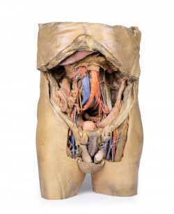
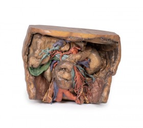
3D anatomical models of the abdominal cavity and internal organs are advanced educational tools that realistically present the anatomical structures of the abdominal cavity, such as bones, muscles, internal organs, blood vessels, lymphatic system and neural structures. Various elements can be identified within the abdominal cavity, including internal organs (small intestine, large intestine, stomach, liver, etc.), blood vessels and structural elements of the abdominal wall. Due to their complexity, these models are extremely valuable in the teaching process, enabling a thorough understanding of the functions and relationships between individual anatomical structures. Our offer includes 3D anatomical models of the abdominal cavity and internal organs, which were created on the basis of radiological data from computed tomography (CT) and using modern 3D printing technology. In addition, we also offer traditional anatomical models of the abdominal cavity.
3D anatomical models of the abdominal cavity and internal organs - advantages:
Our 3D anatomical models of the abdominal cavity and internal organs are made entirely of artificial materials, which means that they do not wear out and provide uniform teaching conditions for all students. An additional advantage of these models is their extremely precise manufacturing, possible thanks to the use of radiological data from CT and 3D printing technology. These are high-fidelity models that precisely reproduce the actual dissection preparation on the basis of which they were created. Our abdominal anatomical models present structures such as bones, muscles, internal organs, nerves, blood and lymphatic vessels in their correct anatomical positions. This is a huge advantage compared to traditional models, which often present anatomy in a more schematic way. Moreover, 3D anatomical models of the abdominal cavity and internal organs do not require special storage conditions and can be used in standard classrooms. Their durability is also very long, which translates into economical use. Undeniably, the matching colors of individual structures make it easier to understand the topography and relationships between them when learning anatomy.
3D anatomical models of the abdominal cavity and internal organs - application:
The 3D anatomical models of the abdominal cavity and internal organs that we present are an ideal educational tool for students of medical faculties such as medicine, physiotherapy, nursing and emergency medical services. Currently, they are widely used at universities around the world. Moreover, these models are perfect for equipping anatomical and biological laboratories and anatomical museums. They are invaluable during courses and training in various fields of medicine. 3D anatomical models of the abdominal cavity and internal organs can also be a decorative element in teaching rooms and medical offices or be part of thematic exhibitions devoted to human anatomy.
Types of 3D anatomical models of the abdominal cavity and internal organs:
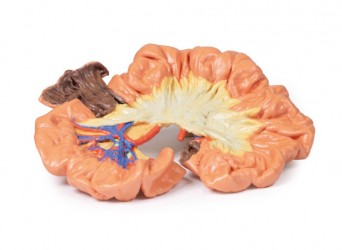
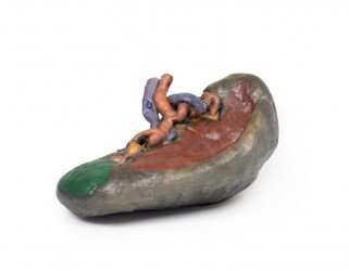
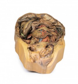
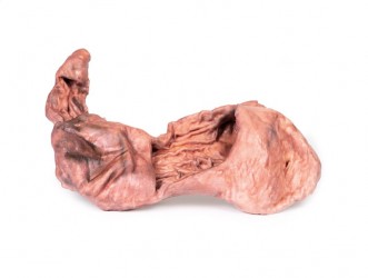
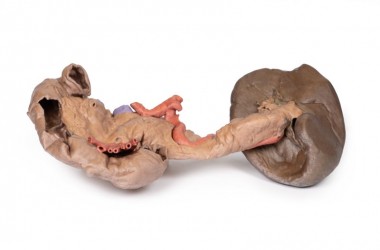
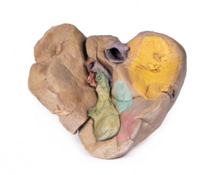
Our offer includes both conventional anatomical models and advanced 3D anatomical models of the abdominal cavity and internal organs, which faithfully reproduce their anatomical structure, just like real dissection preparations. Our offer includes models that allow for a detailed examination of the interior of the abdominal cavity and the location of internal organs. The 3D models that we have in our assortment include: model of the stomach, spleen, pancreas, liver with gallbladder, model of the abdominal cavity with a bilateral hernia.
- The 3D intestinal model is particularly popular.
Based on radiological data, a series of models faithfully reflecting the human body were created. These advanced teaching aids are the equipment of anatomy laboratories of many Medical Universities in Poland. This is a modern solution used in many countries around the world.
How can we help you?
OpenMedis is an experienced manufacturer and distributor of anatomical models on the Polish market. For many years, we have been providing educational tools to anatomy laboratories at medical universities, medical offices, schools and museums. If you need advice on equipment for an anatomy laboratory in your institution, we are ready to provide professional help and advice. Please contact us.
In addition, we also recommend our
3D anatomical models of the head and
3D lung models, which, combined with
3D models of the abdominal cavity and internal organs, create a comprehensive set for learning the anatomy and physiology of the body.


 See our profile on Facebook
 Check our profile on Instagram
 Download a PDF file
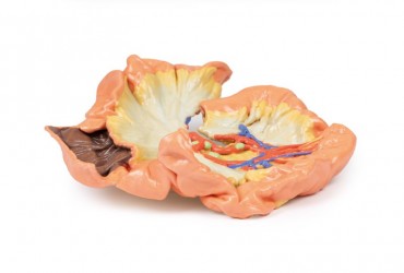

