Clinical History
This elderly patient had a long history of „indigestion“. He collapsed and died after a massive stoke.
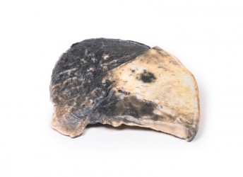
Pathology
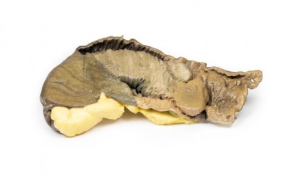
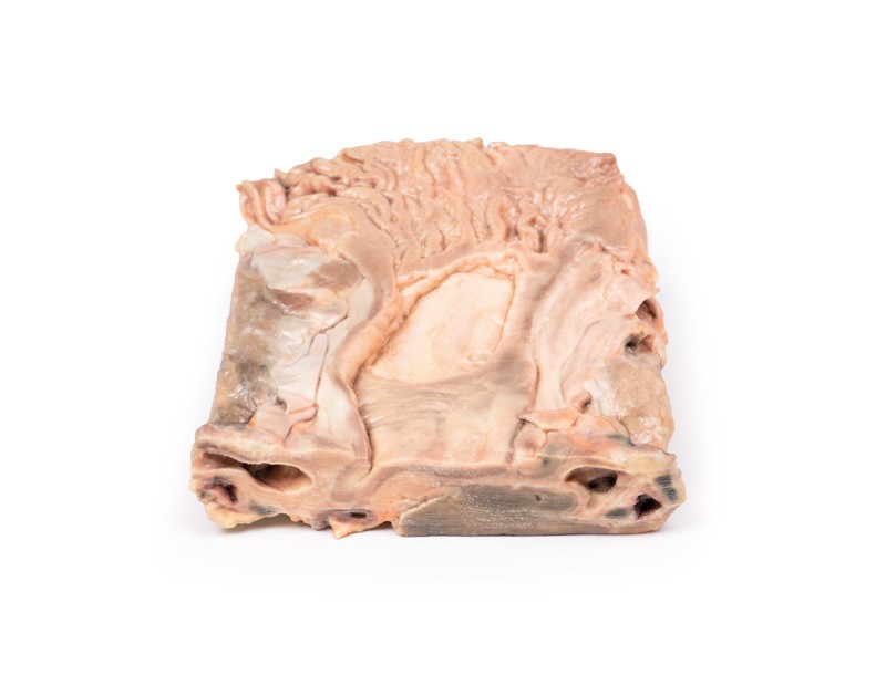
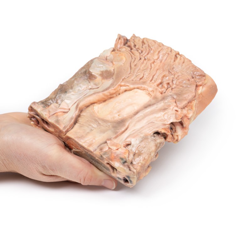
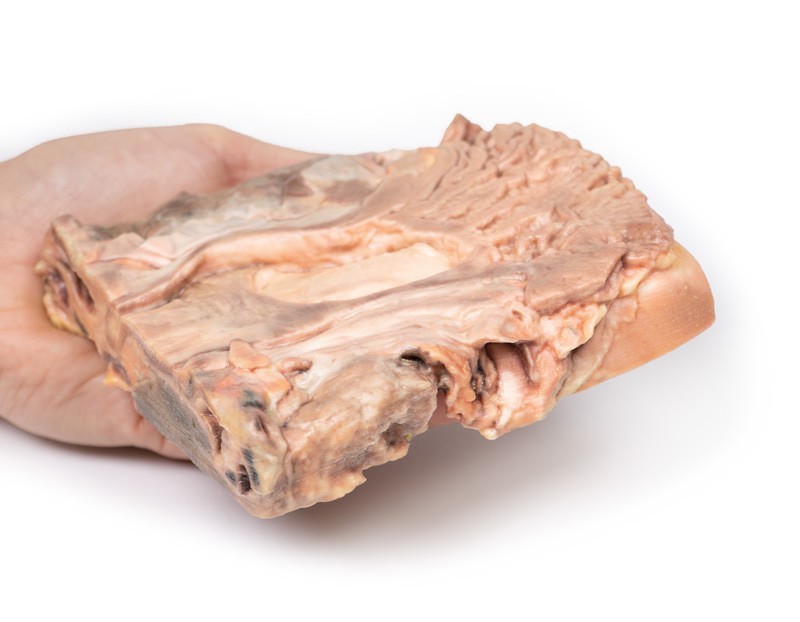
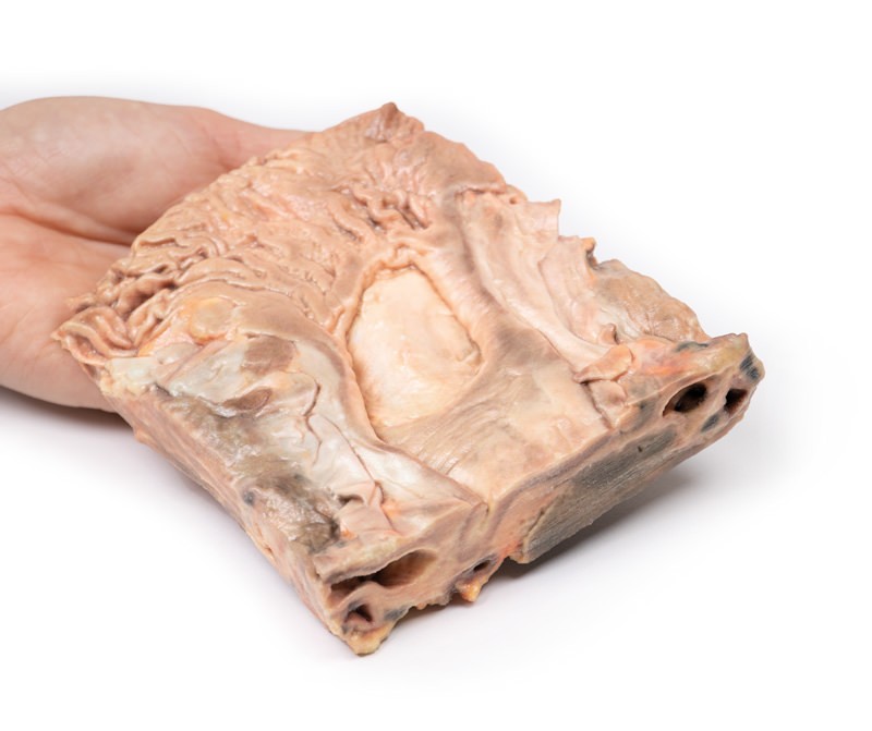
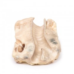
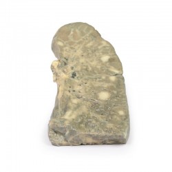
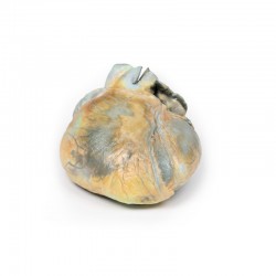
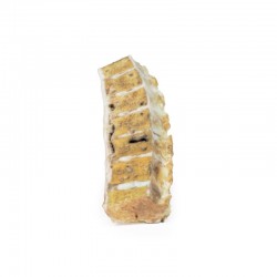
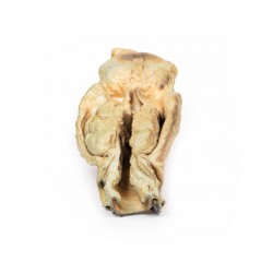
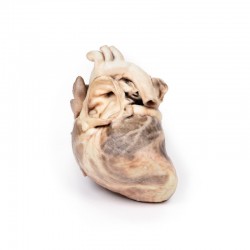
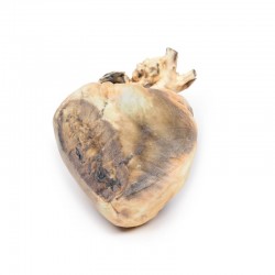
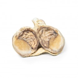
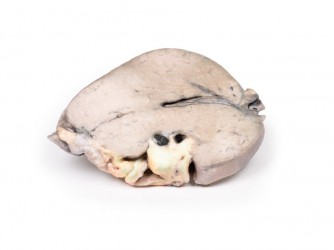
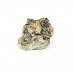
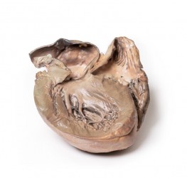
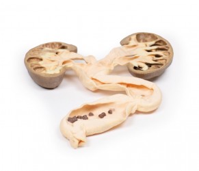
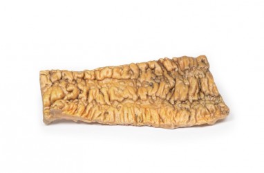
The specimen is a 2cm coronal slice of tissue, which incorporates a portion of stomach diaphragm, liver and pancreas. The specimen has been opened to display a large ulcer at the upper end of the lesser curvature near the gastro-oesophageal junction. Macroscopically, the loss of substance at the site of the ulcer is oval, has 5-6cm in diameter and slightly elevated borders. The base is clean and smooth with no evidence of haemorrhage. The gastric wall surrounding the ulcer is indurated, because of the fibrosis that involves the base of the ulcer and spreads beneath the surrounding mucosa. Being retractile, the fibrosis manages to „pull“ the gastric mucosa towards the base of the ulcer, so that gastric mucosal folds converge radially around the loss of substance (this feature is not seen in ulcerated malignant gastric tumours). This is evident on the inferior aspect of the ulcer and less so superiorly.
Further Information
Patients with a gastric ulcer may experience pain worsening with eating, often described as a burning or dull ache. Other symptoms include belching, vomiting, weight loss, or poor appetite. Complications may include bleeding, perforation, and blockage of the stomach. Common causes include the bacteria Helicobacter pylori and non-steroidal anti-inflammatory drugs (NSAIDs). H. pylori was first identified as causing peptic ulcers by Barry Marshall and Robin Warren of the University of Western Australia in the late 20th century, a discovery for which they were awarded the Nobel Prize in 2005. Other, less common causes include tobacco smoking, stress due to serious illness, Beh et’s disease, Zollinger-Ellison syndrome, Crohn’s disease, and liver cirrhosis[1]. Older people are more sensitive to the ulcer-causing effects of NSAIDs[1]. The diagnosis is typically suspected due to the presenting symptoms with confirmation by either endoscopy or barium swallow[1]. H. pylori can be diagnosed by testing the blood for antibodies, a urea breath test, testing the stool for signs of the bacteria, or a biopsy of the stomach[1]. Other conditions that produce similar symptoms, include stomach cancer, coronary heart disease, and inflammation of the stomach lining (gastritis) or gallbladder inflammation (cystitis) [1]. Treatment includes stopping smoking, stopping use of NSAIDs, reducing or preferably stopping alcohol consumption, and taking medications to decrease stomach acid[1]. Ulcers due to H. pylori are treated with a combination of medications, such as amoxicillin, clarithromycin, and a proton pump inhibitor (PPI). The medication used to decrease acid is usually either a PPI or an H2 blocker (histamine H2-receptor antagonists). Bleeding ulcers may be treated by endoscopy, with open surgery typically only used in cases in which it is not successful. Peptic ulcers are present in around 4% of the population.


 Download a PDF file Add to quotation - wish list
