


Home / 3D anatomy models / 3D anatomical models of pathologies and diseases / Hepatic duct calculi and Obstructive Biliary Cirrhosis
Hepatic duct calculi and Obstructive Biliary Cirrhosis

Hepatic duct calculi and Obstructive Biliary Cirrhosis
 Download a PDF file Add to quotation - wish list
Product description: Hepatic duct calculi and Obstructive Biliary Cirrhosis

Clinical History
An 85-year old male presented with urinary retention due to benign prostatic hypertrophy. On admission it was noted that he was jaundiced with cholestatic derangement of his liver function tests. He underwent a transurethral prostate resection but died from pneumonia 5 days post-operative.
Pathology
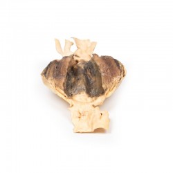
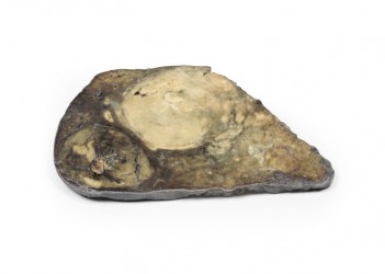
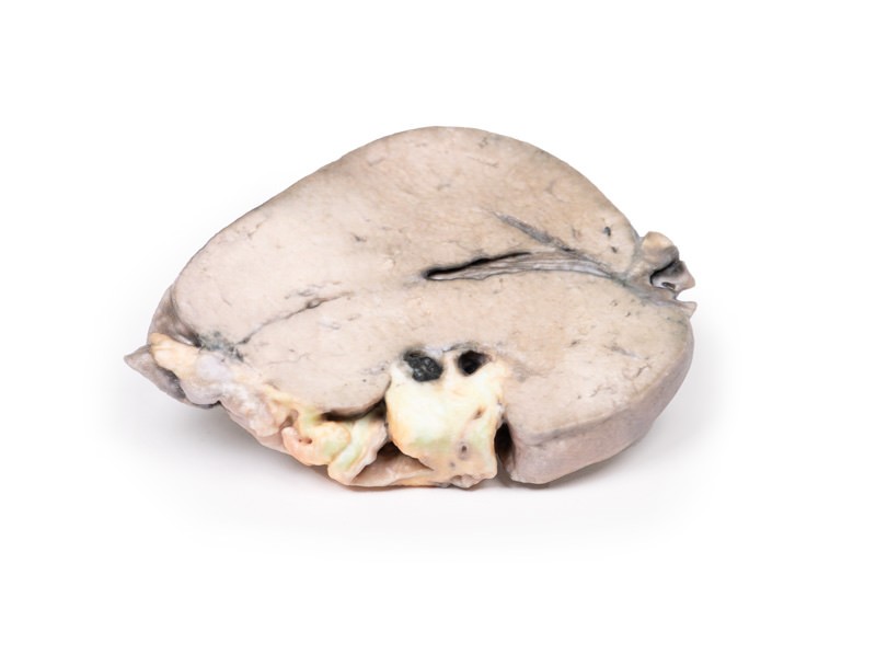
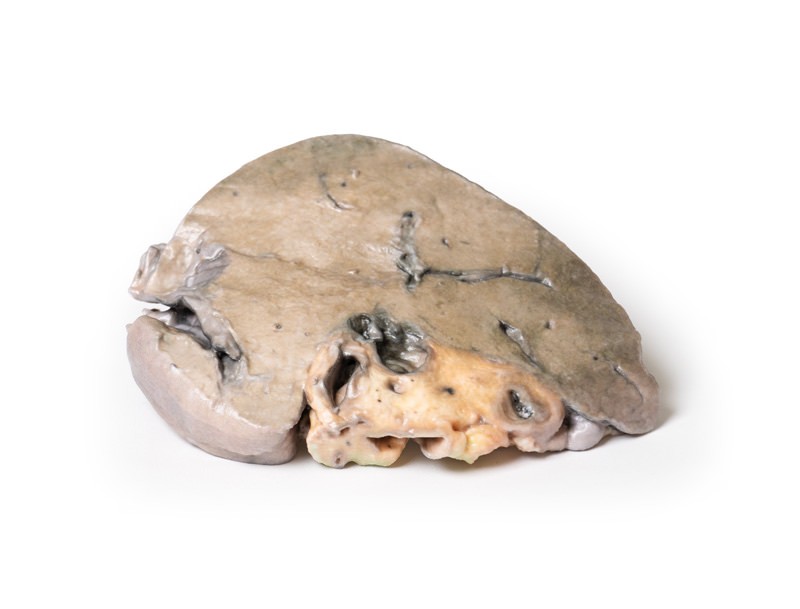
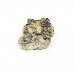
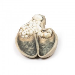
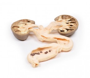
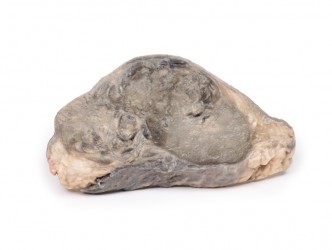
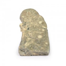
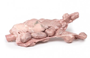
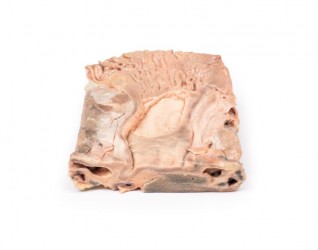
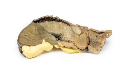
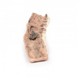
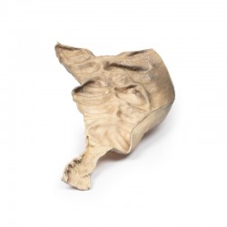
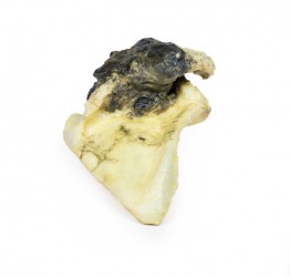
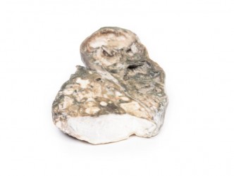
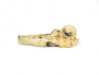
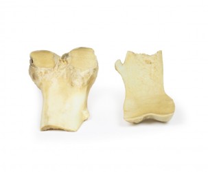
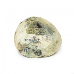
The specimen is a slice of liver mounted to display the cut surface. The capsule is slightly thickened and the liver substance has a finely nodular appearance. Intrahepatic bile ducts are dilated. When the posterior or inferior surface is viewed an irregular pigmented calculus, 10 mm in diameter, is seen impacted in a distended hepatic duct. Another smaller calculus 3 mm in diameter has been dislodged. This specimen represents an example of secondary biliary cirrhosis due to large duct obstruction from hepatic calculi.
Further Information
Hepatolithiasis is characterised by the presence of intrahepatic gallstones. These calculi can lead to cholangitis, progressive hepatocyte atrophy and destruction, and an increased risk of cholangiocarcinoma. It is common in East Asia but rare in Western countries. There is no difference in incidence between genders. The stones are most commonly made up of pigmented calcium bilirubinate stones.
These stones cause intrahepatic bile duct obstruction. Proximal to the obstructing stone distension and dilation of the bile ducts is evident. There is also bile duct proliferation at the portalparenchymal interface with stromal oedema and infiltrating neutrophils, indicating an acute-chronic inflammation. If untreated this inflammation leads to periportal fibrosis and eventually obstructive biliary cirrhosis. Microscopic appearance would show feathery degeneration of periportal hepatocytes, cytoplasmic swelling often with Mallory Denk bodies (i.e. an inclusion found in the cytoplasm of liver cells with twisted-rope appearance caused by damaged intermediate filaments within the hepatocytes) and bile infarcts from extravasated bile. Chronic inflammation can lead to biliary dysplasia which may develop into cholangiocarcinoma. Patients may present with repeated cholangitis, intermittent abdominal pain, jaundice or frequently no symptoms. Treatment is usually surgical removal of the calculi.
Inquiry
Related products
