Quality Certyficate
street: Kolejowa 2, 30-805 Cracow


Home / Anatomical Models / Upper extremities models / Hand model
Anatomical Models
3D anatomy models
Custom tools for patient education
Veterinary simulators
Anatomical Charts
Anatomical Table 3D
Medical simulators
Medical Equipment
Type:
 See our profile on Facebook
 Check our profile on Instagram
 Download a PDF file
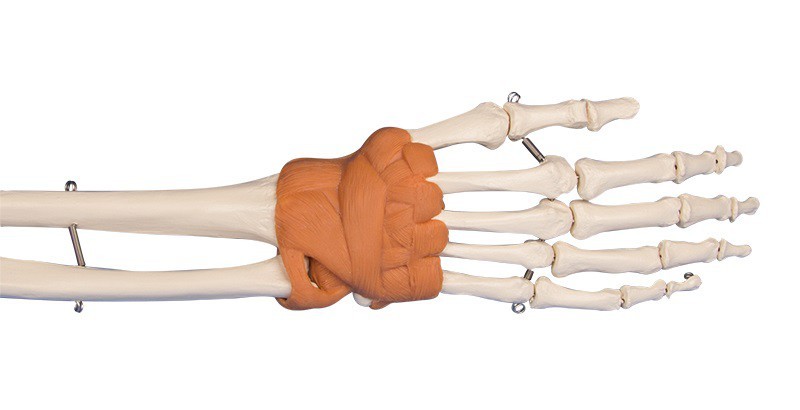
Anatomical Models / Upper extremities models

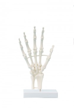
Hand and lower arm with representation of the wrist ligaments.
All bones are individually moulded and mounted on wire. Life size.
...
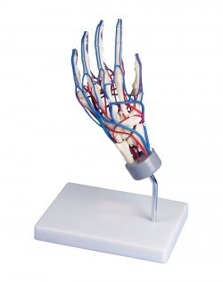
A life-size model showing hand vasculature. Main veins and arteries are shown. The circulatory system is visible on both the dorsal and palm sides of the hand.
Additional information:
Size 26 x 15 x 22 cm
...
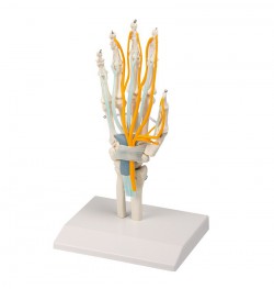
Hand with start of lower arm, with Tendons, Carpal Tunnel and Nerves. The skeleton is mounted on wire and is composed of single bones. The model shows the following structures: tendons of flexor digitorum ...
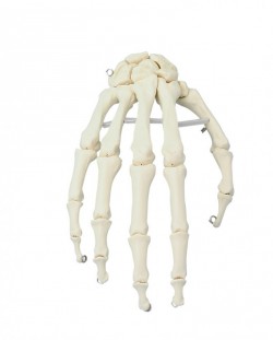
A life-size anatomical model showing the skeletal structure of the hand. It Shows all palm bones which are individually mobile-mounted on wire
It is possible to perform translational movements in individual hand ...
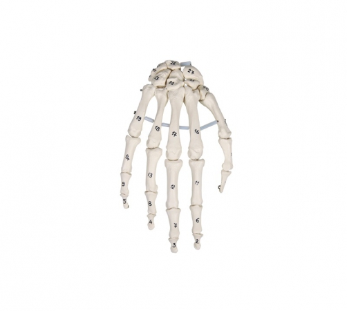
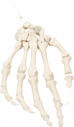
True to life casting of a skeleton of the human hand. All hand bones are individually mobile-mounted on wire. With additional numbering of the individual hand bones. ...
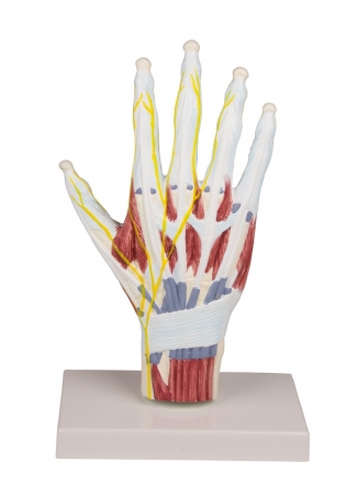
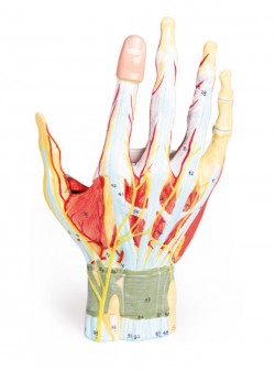
Life-size anatomical model of the hand. It presents hand-made muscles of the hands, ligaments, nerves, bones and tendons. Made on the basis of. There is a possibility remove the model from the base.
Additional ...
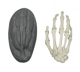
Natural, non-movable one-piece casting of a human hand. Representation of all structures and anatomical details. ...
This hand skeleton is especially suitable for those who wish to examine the bones in detail. Due to the loose nylon mount, the bones can be separated and viewed individually. Nevertheless, they always remain in the hand ...
The anatomical model of the right human hand is an ideal teaching aid for medical students who want to explore the details of the anatomical structure of the hand. The model is made entirely of artificial material, ...
An innovative concept that adds a dynamic element to the study of the anatomy of the hand. Strategically placed magnets help learners understand the complex structures of finger bones and their joints. All bones in this ...
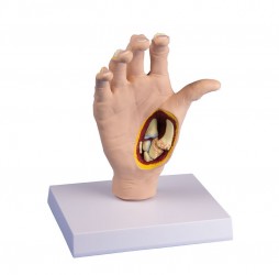
Life-size right hand with cross-section showing the effects of osteoarthritis, including osteophytes (bone processes), Heberden and Bouchard nodes, and thumb swan deformity. Other anatomy affected by osteoarthritis is ...
1
Anatomical Models - Upper extremities models
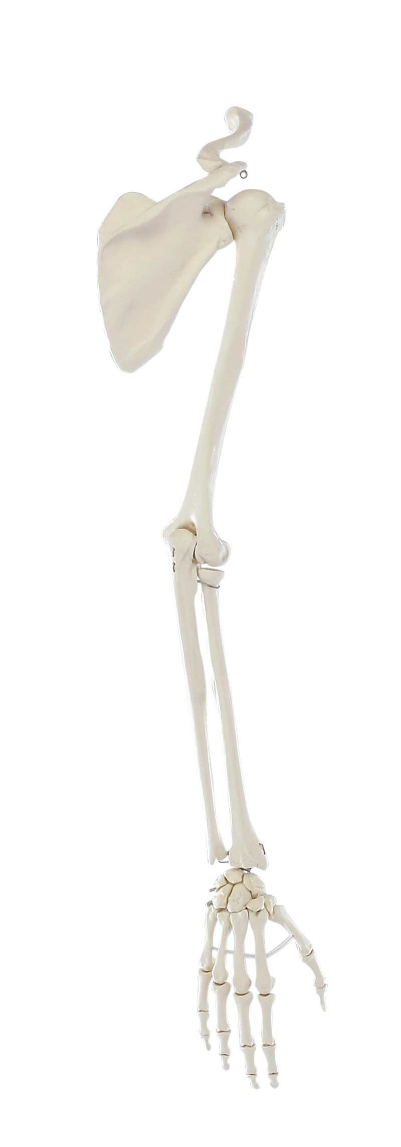
The human upper limb is divided into the shoulder girdle, forearm and arm (hand). The shoulder girdle is made up of the scapula, clavicle and the proximal part of the humerus. The forearm consists of two long bones: the ulna and the radius. The hand is a complex system of bone connections, which includes the bones of the wrist, metacarpals and phalanges. Anatomical models facilitate education in the field of osteology of the upper limb and the biomechanics of its joints, i.e. shoulder joint, elbow joint, proximal and distal radioulnar joint, radiocarpal joint, metacarpo-digital joints and proximal and distal interphalangeal joints.
What does the OpenMedis online store offer?
The offer includes teaching aids presenting important elements of the anatomical structure of the upper limb and the functioning of the joints located within it. The functional connection of individual joint elements - using an elastic rubber, allows the presentation of anatomical and translational mobility. The most popular product, which is particularly popular among doctors, physiotherapists and manual therapists, is the upper limb model with a flexible shoulder girdle. Thanks to such precise work, learning anatomy becomes even more effective.
Depending on your needs, we can also offer an anatomical model of the upper limb with manually applied muscle attachments.
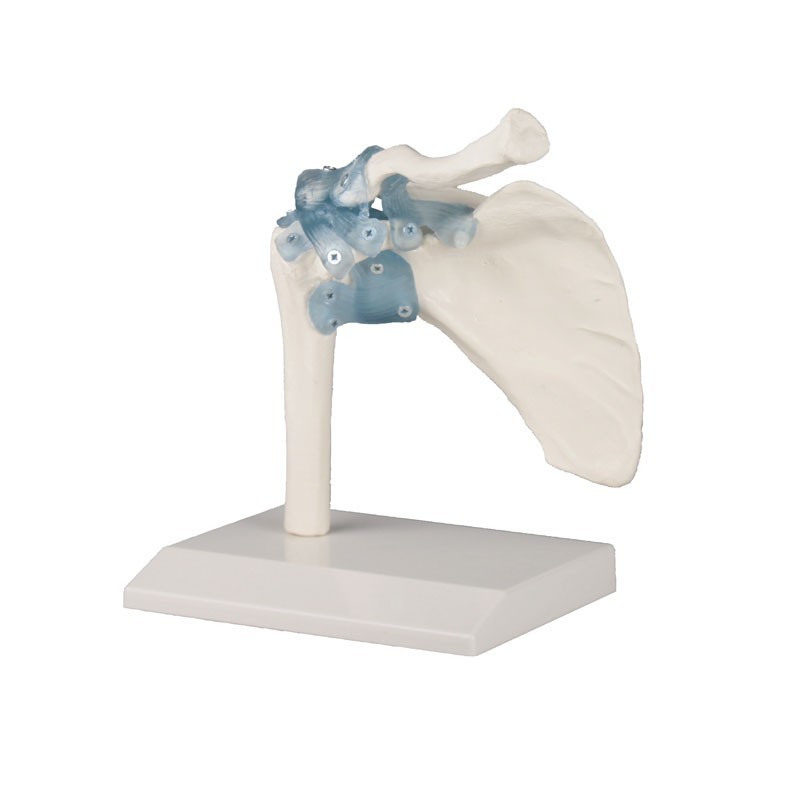
Our offer also includes anatomical models of individual joints of the upper limb: e.g. a shoulder joint model, an elbow joint model, a forearm model with a radiocarpal joint.
The basic and simplest product in this category is a life-size bone model of the hand showing the bone topography, mutual relations between the bones and the structure of the hand joints. Its modifications include an anatomical model of the hand with numbered bone elements. The anatomical model of the hand with tendons, nerves and carpal tunnel is a life-size cast model with distal fragments of the forearm bones, schematically presented tendons and nerves running in the carpal tunnel. The 7-piece hand model deserves special attention. It presents the anatomy of the hand with great accuracy and detail. The superficial layers can be removed at various levels to reveal the internal structures. The model shows muscles, blood vessels, nerves, tendons, ligaments and bones. The most detailed 3D anatomical model of the hand shows muscles, tendons, vessels and nerves.
Our offer also includes vascular models. These are a life-size vascular anatomical model of the hand and a comprehensive model of the upper limb with blood vessels.
OpenMedis offer:
- Model of the upper limb with a flexible shoulder girdle
- Upper limb model with shoulder girdle and muscle markings
- Anatomical model of the hand and wrist
- Vascular model of the hand
- Model of the upper limb with blood vessels
- Model of the shoulder joint
- Model of the elbow joint
- Model of a hand with tendons, nerves and carpal tunnel
- Model of the shoulder joint with deep muscles
- Model of the shoulder joint with muscles
- Hand anatomy, 7 parts
- Arm muscles model
Advantages of anatomical models of the human upper limb:
- Accuracy of workmanship (hand-finished)
- Durability (made of the highest quality materials)
- Flexible joint connections to demonstrate translational and anatomical mobility
- Learning the anatomy of the normal human upper limb
- Learning the topography of the anatomical structures of the upper limb and the mutual relations between them
Application of the human upper limb model:
- Equipment of the anatomy laboratory
- Learning the anatomy of the human upper limb region
- The anatomical model can be used to demonstrate the structure of the upper limb and its injuries/pathologies - to patients in an office setting
- As a teaching aid during postgraduate training for doctors, physiotherapists, osteopaths and other medical professions.
Anatomical models of the human upper limb are ideal teaching aids for everyone who wants to deepen their knowledge about the anatomical structure of the upper limb and the biomechanics of its joints. Using different colors to paint individual elements makes it easier to learn the topography of anatomical structures and the mutual relations between them. Thanks to the use of the highest quality materials and advanced production methods, models of the human upper limb are characterized by high durability and precision. Many anatomical models of the upper limb, presenting the bone skeleton, are real casts made of real human bones. Our assortment includes both bone models of the human upper limb and anatomical models showing the vascularization and innervation of the upper limb or musculature. Depending on the type, anatomical models of the human upper limb present the skeleton of the upper limb as a whole, the skeleton of the upper limb in fragments, the structures of the hand region, and the course of blood vessels and nerves. Some anatomical models can be broken down into individual elements (e.g. a model of the muscles of the upper limb), which makes learning anatomy much more attractive and easier. Hand-made markings of muscle attachments also bring great educational value. These types of anatomical models are delivered with an educational card containing the nomenclature of anatomical structures of the upper limb.
The human upper limb model is ideal for learning anatomy at home and during seminars conducted at universities and medical schools. Based on radiological data, a series of models faithfully reflecting the human body were created. These advanced teaching aids are the equipment of anatomy laboratories of many Medical Universities in Poland. This is a modern solution used in many countries around the world.
Our anatomical models of the upper limbs are made with full attention to detail. They allow for accurate imaging of real anatomical structures. They provide support for health education, research and practice.
We also recommend our models of the skull, spine and lower limbs, which, combined with anatomical models of the upper limbs, will create an ideal set for learning anatomy.
If you have any questions or if the upper limb model you need is not available in the store, please contact us via e-mail or telephone. We are sure that together we will be able to provide the necessary materials for learning and improving skills. We can produce models to order.
