Medical simulators / X-ray/CT Phantoms
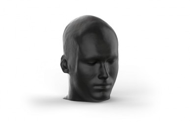
Human skull designed for radiographic examinations, embedded in a special, transparent material that does not cause image artifacts.
Main features:
The jaws are slightly open to allow dental panoramic ...
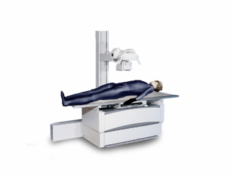
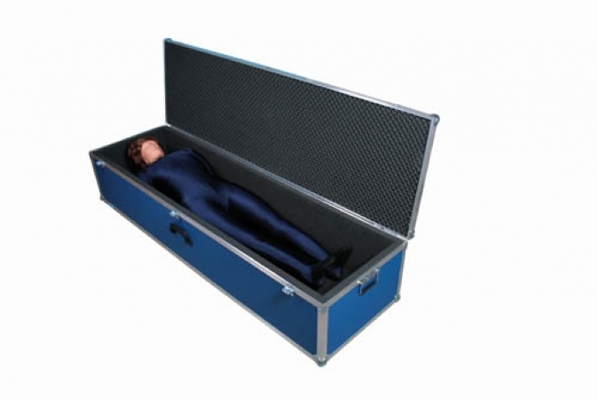
Life-size X-ray Phantom. It contains a real human skeleton that allows taking real X-ray images comparable to a real patient. Using a real skeleton allows to visualize even the smallest anatomic details, which is ...
This model offers all features of model SM01133 but includes a plastic skeleton and is due to this only suitable for positioning training. ...
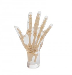
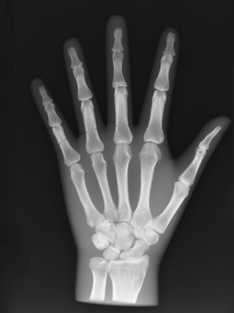
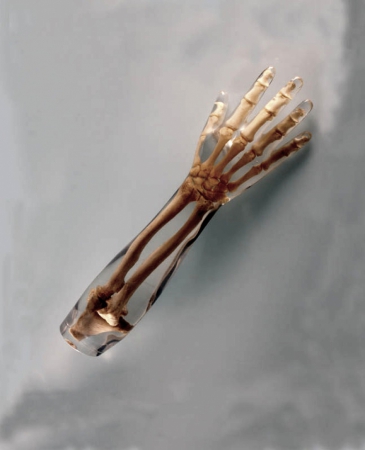
An adult man's hand together with wrist bones and distal fragments of the forearm bone, embedded in a special, transparent material that does not cause image artifacts. Phantom is designed to take X-ray pictures.
Main ...
An adult man's hand together with wrist bones and distal fragments of the forearm bone, embedded in a special, non- transparent material that does not cause image artifacts. Phantom is designed to take X-ray ...
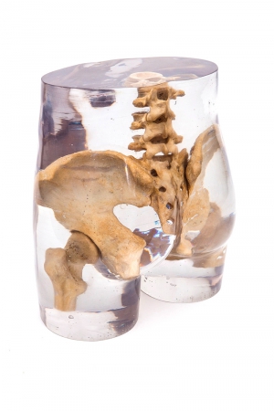
The pelvis of an adult human with the lower lumbar vertebrae and proximal parts of the femurs, embedded in a special, transparent material that does not cause image artifacts. Phantom is designed to take X-ray ...
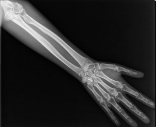
Phantom containing the bones of the forearm, wrist, mediastinum and phalanges, intended for performing X-ray photography.
The bones were embedded in a special, transparent material that did not cause image ...
Phantom containing the bones of the forearm, wrist, mediastinum and phalanges, intended for performing X-ray photography.
The bones were embedded in a special, non-transparent material that did not cause image ...
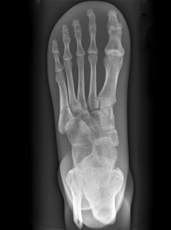
Phantom containing the bones of the foot and distal part of tibia and fibula, intended for performing X-ray photography.
The bones were embedded in a special, transparent material that did not cause image ...
Phantom containing the bones of the foot and distal par of tibia and fibula, intended for performing X-ray images.
The bones were embedded in a special,non- transparent material that did not cause image ...
The pelvis of an adult human with the lower lumbar vertebrae and proximal parts of the femurs, embedded in a special, non-transparent material that does not cause image artifacts. Phantom is designed to take X-ray ...
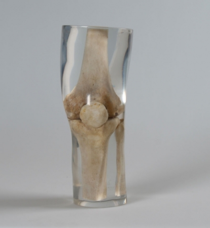
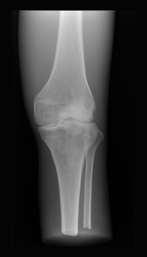
A phantom of the knee joint designed for performing X-ray photography. Has a distal part of the femur, proximal fragment of tibia and fibula bone and patella. The whole is embedded in a special, transparent material, ...
A phantom of the knee joint designed for performing X-ray photography. Has a distal part of the femur, proximal fragment of tibia and fibula bone and patella. The whole is embedded in a special, nontransparent ...
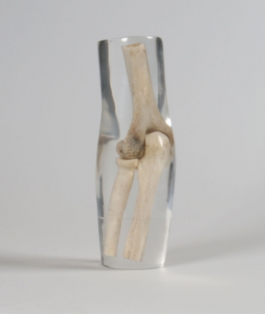
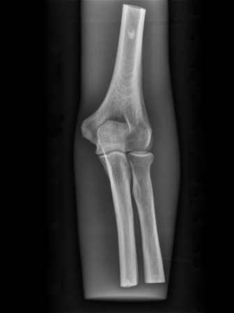
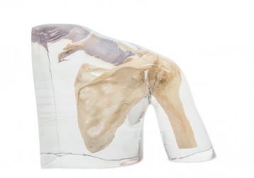
An adult man's elbow joint, embedded in a special, transparent material that does not cause image artifacts. Phantom is designed to take X-ray pictures.
Main features:
Phantom has a humerus, radius and ...
This X-Ray part phantom gives the unique opportunity to take x-ray images of single body parts again and again. The Phantom includes real human bones and allows taking real x-ray images. The model is perfect for ...
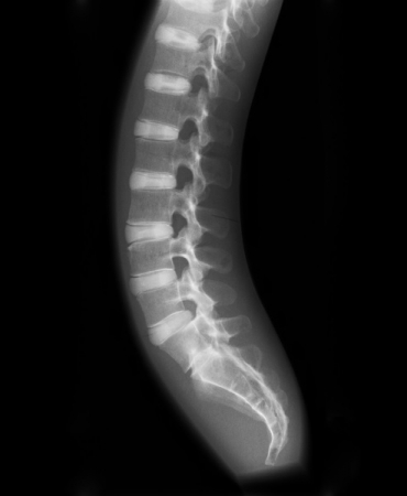
A vertebral column phantom designed for performing X-ray photography. The following bones were embedded in the nontransparent material:
Cervical, thorax, lumbar, sacral vertebrae
Sacrum
Main ...
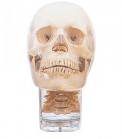
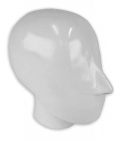
Human skull designed for radiographic examinations, embedded in a special, transparent material that does not cause image artifacts.
Main features:
The jaws are slightly open to allow dental panoramic images ...
Human skull designed for radiographic examinations, embedded in a special, non-translucent material that does not cause image artifacts.
Main features:
The jaws are slightly open to allow dental panoramic ...
A chest phantom designed for performing X-ray photography. The following bones were embedded in the soft material:
vertebrae from C6 to L3,
ribs,
the shoulder blades
collarbones.
Main ...
This partial x-ray phantom offers the unique ability to repeat actual x-rays as often as you like. The model contains real human bones, making it possible to take real photos. The model is perfect for schools and ...
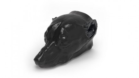
Phantom provides a realistic simulation of a dog's head without a contrast agent. Bones and soft tissues are displayed authentically with realistic CT values for all tissues at a lamp voltage of 120 kWp in CT. The air ...
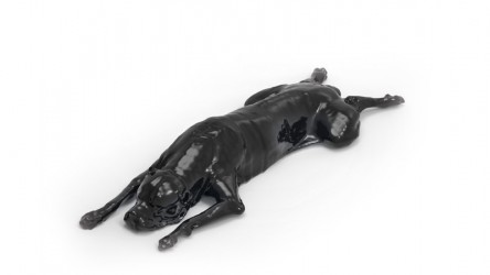
Phantom provides a realistic simulation of a dog without a contrast agent. Bones and soft tissues are displayed authentically with realistic CT values for all tissues at a lamp voltage of 120 kWp in CT. The air spaces ...
Breast phantom with adipose and glandular tissue. This breast phantom was developed to simulate breast imaging in mammography and tomosynthesis.
It shows a breast 4 cm thick, which can be fixed under a ...
This phantom is created from real patient data and is produced using the latest technology. Bones, vessels and soft tissues are displayed authentically with realistic CT values for all tissues at a lamp voltage of 120 ...
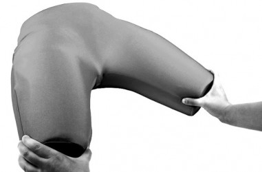
The flexible hip x-ray phantom contains real human bones and offers the unique opportunity to take real x-rays. The phantom consists of the entire pelvis, including the coccyx, the two lumbar vertebrae (L4 + L5), and ...
This phantom is created from real patient data and is manufactured using the latest technology. Bones, vessels and soft tissue are displayed authentically with realistic CT values ??for all tissues at 120 kVp tube ...
This phantom is created from real patient data and is manufactured using the latest technology. Bones, vessels and soft tissue are displayed authentically with realistic CT values for all tissues at 120 kVp tube voltage ...
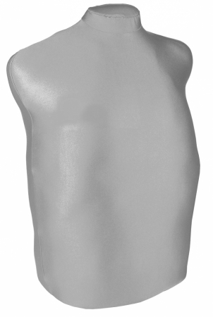
The advanced ultrasound trainer is the anatomy model of an adult human torso for ultrasound examination, including the FAST protocol. The torso phantom for ultrasound examinations has an actual size with ...
1
Medical simulators - X-ray/CT Phantoms
Radiological phantoms are extremely advanced devices that reflect real and realistic clinical conditions, without the participation of actual patients. These devices allow you to practice and improve medical skills in the field X-ray/CT. Medical simulation is one of the popular education methods using specialized educational equipment - from trainers with a simple structure and functions, intended for learning simple, single medical procedures and tasks, through advanced phantoms, mannequins, simulators with a more complex structure - in the form of complete, e.g. whole patients faithfully reflecting the parameters and structure of real people. The main goal of medical simulation is to prepare future medical staff to work with real patients by creating the most optimal, safe and real training conditions that faithfully reflect the actual situation. The undoubted advantage of medical simulation is the shortening of the time required for proper preparation for the profession compared to traditional teaching methods. Medical simulators are one of the most necessary educational tools for students of various medical studies - doctors, nurses, paramedics, physiotherapists. Medical radiological trainers can be used by students as a training tool for practical skills acquired during theoretical lectures. Thanks to them, the risk associated with inappropriate performance of a specific activity is reduced by one hundred percent.
Radiological phantoms:
Radiological phantoms, advanced models for simulating medical imaging procedures, include comprehensive phantoms, dedicated CT, X-ray, as well as models representing specific body parts.
Radiological phantoms are used in various areas, such as quality control and device calibration, dosimetry and education in the area of proper positioning.
- Radiological phantoms - anthropomorphic - anthropomorphic radiological phantoms are models that imitate the human body, made of materials with similar properties to the tissues of biological organisms. In contrast to the need to examine real patients, anthropomorphic phantoms allow for trials and experiments that help assess the optimal use of radiation, for example in new protocols or image reconstruction techniques. In addition, these radiological models are also used to train medical staff in the correct positioning of patients and taking accurate diagnostic images. Anthropomorphic radiological phantoms can be used to teach various imaging techniques and factors related to radiation exposure.
- Radiological phantoms - calibration - radiological phantoms that perform a calibration function are usually cylinders or plates made of materials with precisely known densities. They are used to monitor the quality of medical images, ensuring that phantom image reconstructions reflect the correct density values. If deviations from these known values are noticeable, it may indicate the need for maintenance or repair of the imaging equipment.
Whole radiological phantoms are comprehensive models that reproduce the anatomical structures of the body. They are used to practice diagnostic procedures, improving the skills of medical staff and students, without the need to expose patients to radiation.
Radiological phantoms representing specific parts of the body enable focused training in specific diagnostic areas such as the head, brain, lower and upper limbs.
Radiological phantoms are characterized by advanced precision, enabling realistic simulation of clinical conditions and improving the skills of medical teams. They are a key tool in the educational process, contributing to raising the standards of health care and patient safety.
Thanks to current scientific research and constant monitoring of progress in the field of medicine, we are able to adapt our radiological models to the individual needs of the client, including specific pathologies and anatomical anomalies of internal body structures. Our ability to adapt to the changing demands of medicine allows us to deliver tools that meet the highest standards of quality and accuracy in the field of X-ray/CT.
Radiological phantoms - OpenMedis offer:
Our online store offers a wide selection of X-ray/CT Phantoms. Below we present the most frequently selected models from our offer:
We encourage you to check out the full offer of radiological phantoms on our website.
Radiological phantoms - application:
- equipment for medical facilities and hospitals
- equipment for laboratories at universities as demonstration and training equipment
- training equipment during training and courses in the field of medical issues
We recommend that you familiarize yourself with the full offer of medical simulators for learning X-ray/CT. Thanks to the wide range of our online store and the possibility of contact by phone and e-mail, you can easily assemble the appropriate set of medical devices and equipment that you need.
If you have any questions or if the medical simulator for medical imaging you need is not available in the store, please contact us via e-mail or telephone.


 See our profile on Facebook
 Check our profile on Instagram
 Download a PDF file
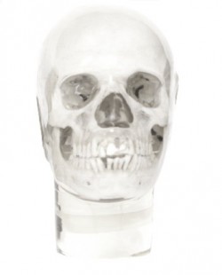


