


Home / 3D anatomy models / 3D anatomical models of pathologies and diseases / Osteosarcoma of femur
Osteosarcoma of femur
Osteosarcoma of femur
 Download a PDF file Add to quotation - wish list
Product description: Osteosarcoma of femur

Clinical History
On examination, there was a palpable tender swelling above the right knee. Blood test showed a raised Alkaline Phosphatase level. A knee x-ray showed periosteal reactive changes in the distal femur suspicious for a bone malignancy. The patient then underwent staging CT and MRI evaluation of the right leg. He underwent adjuvant chemotherapy prior to resection of his right femur. He made a full recovery.
Pathology
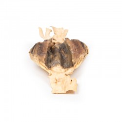
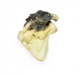
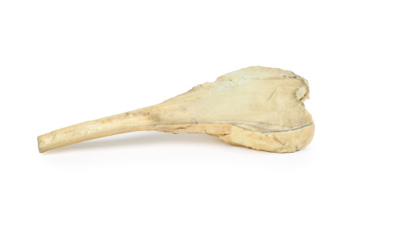
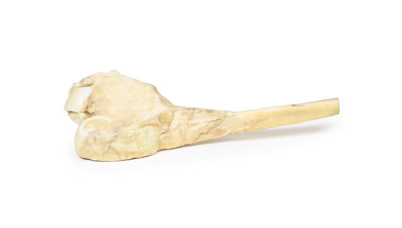
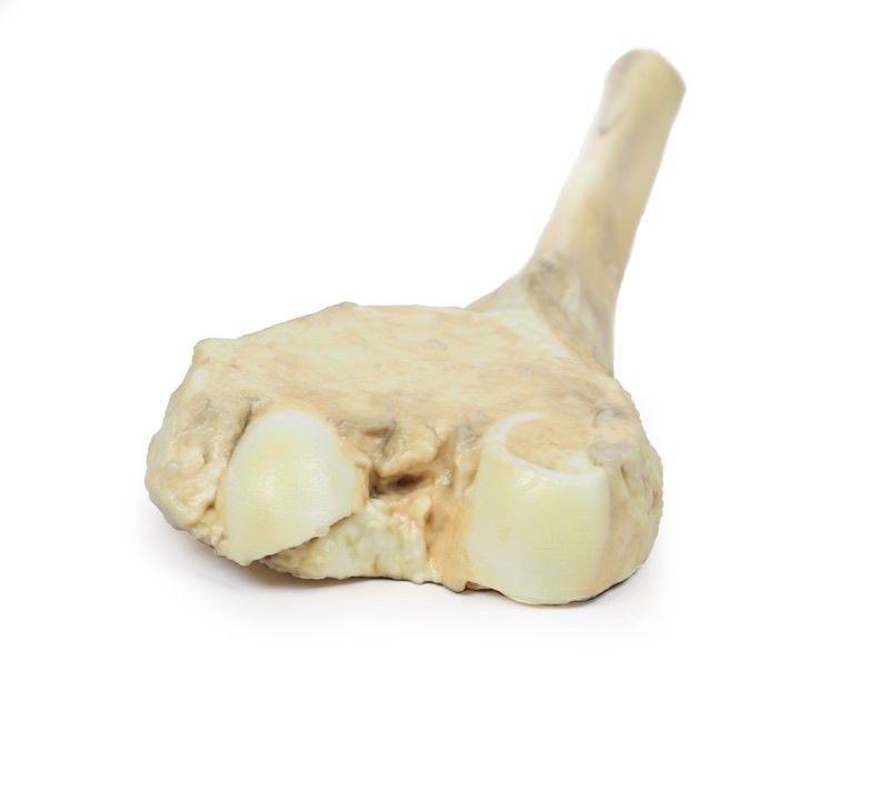
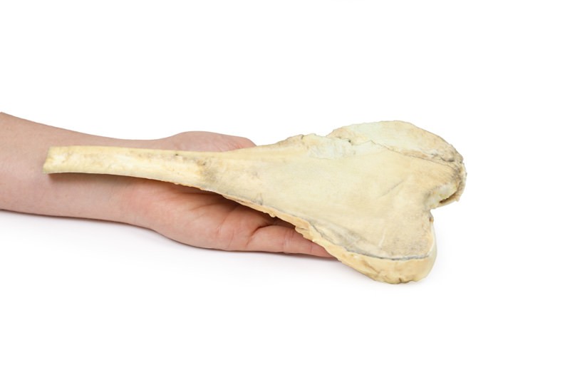
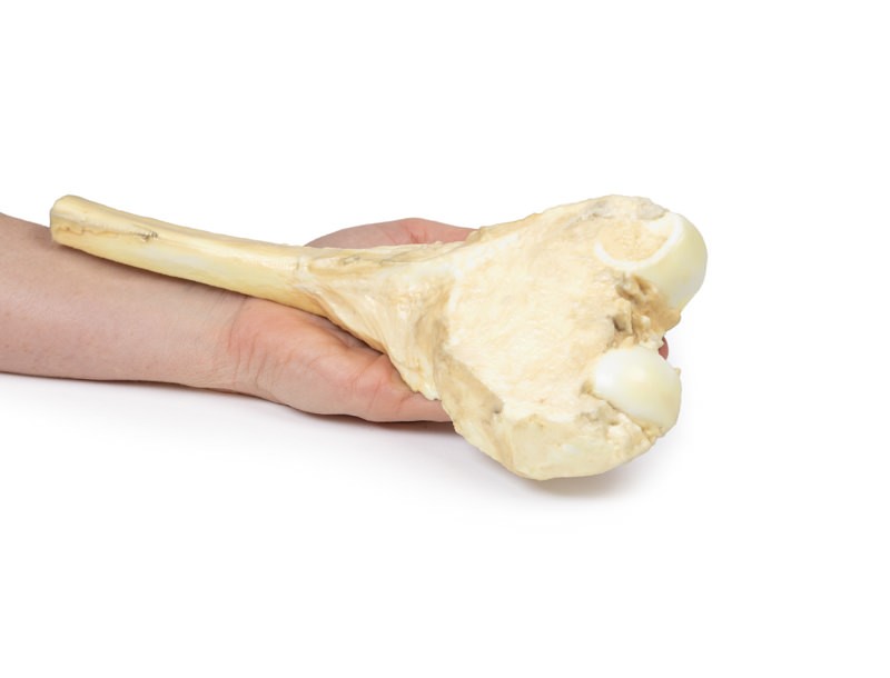
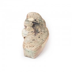
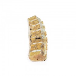
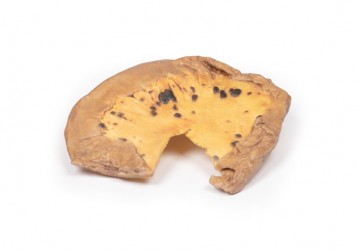
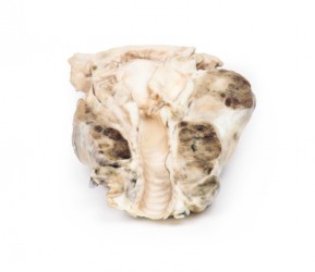
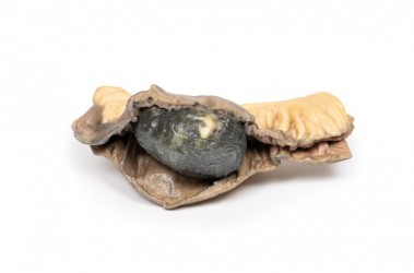
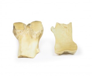
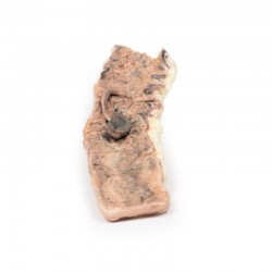
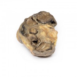
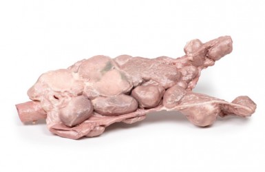
The specimen is the patient’s excised distal femur. On the cut surface, there is a large pale infiltrating tumour, 10 cm in greatest diameter, extending through the periosteum near the articular surface. This is an osteosarcoma of the femur.
Further Information
Osteosarcomas are a malignant tumour of bones that are characterised by the production of osteoid matrix or immature bone. It is the most common primary malignancy of bone. Most occur in the distal femur with the tibia and humerus being the most frequent sites affected. Men are more commonly affected than women. They occur in a bimodal age distribution, with most occurring in children and adolescents under 20 years of age and the second peak occurring in older adults over 60.
Secondary osteosarcomas are more common in older patients. Secondary osteosarcomas occur in patient’s bones with predisposing conditions such as Paget‘s disease, bone infarcts and previous irradiation. Mutations in tumour suppressors and oncogenes, such as RB, TP53 and INK4a have been shown in osteosarcomas.
Osteosarcomas usually present with painful, enlarging masses. Pathological fractures can also be the first presenting complaint. Constitutional symptoms are usually not present. Alkaline phosphatase and lactate dehydrogenase may be elevated on blood tests. X-rays can show features of bone destruction, a mass or signs of a periosteal reaction, such as a sunburst appearance or triangular shells of reactive bone (Codman‘s Triangle). MRI of the affected bone is used to evaluate local staging of the tumour while CT of the body is used to evaluate for distant spread. The tumour may be biopsied in some cases.
The lungs are the most common site for distant metastases followed by the bones and brain. Treatment involves neoadjuvant chemotherapy followed by surgery. 5-year survival rate for localised osteosarcoma is 60-70% but this drops to <20% in patients with distant metastases.
Inquiry
Related products


