


Home / 3D anatomy models / 3D lung anatomical models / Lobar pneumonia - Grey Hepatisation Phase
Lobar pneumonia - Grey Hepatisation Phase
Lobar pneumonia - Grey Hepatisation Phase
 Download a PDF file Add to quotation - wish list
Product description: Lobar pneumonia - Grey Hepatisation Phase

Pathology
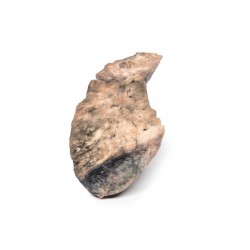
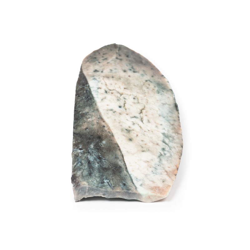
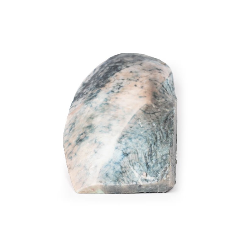
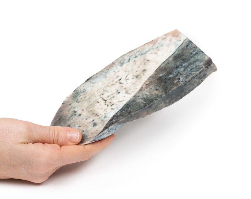
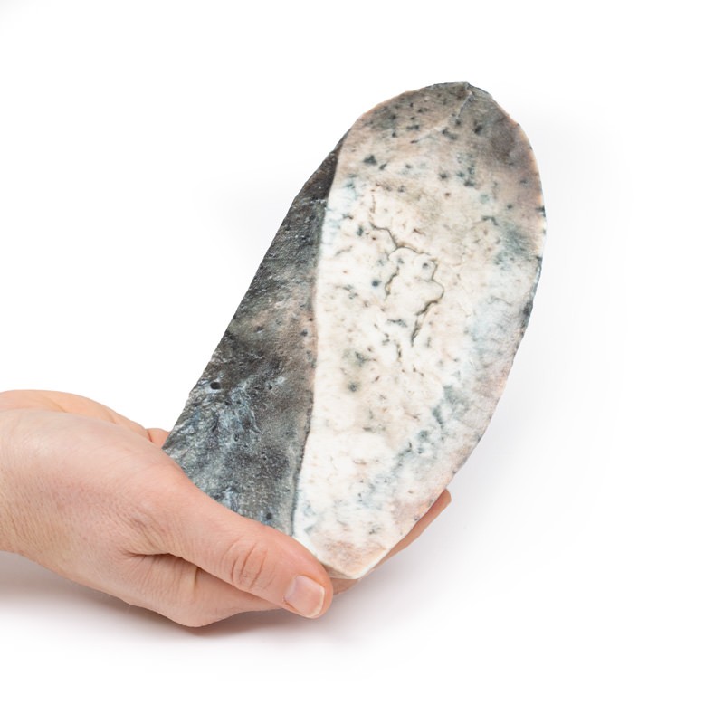
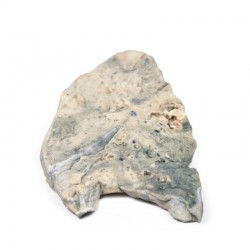
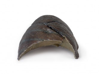
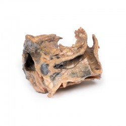
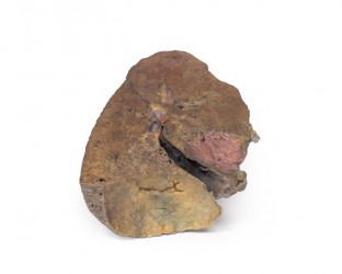
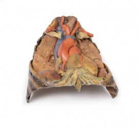
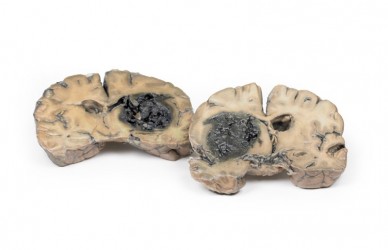
The specimen is a parasagittal section of the right lung and the boundaries between the upper and lower lobes is clearly visible. The entire upper lobe is congested and pale grey in colour.
Further Information
This is an example of a stage of lobar pneumonia in which the inflammatory exudates within the intra-alveolar space resulting in consolidation that affects a large and continuous area of the lobe of a lung. The affected lobe in this case shows grey hepatisation or late consolidation. This usually occurs 2 to 3 days following red hepatisation, and lasts for 4 to 8 days. The lung appears grey with liver-like solid consistency, due to a fibrinopurulent exudate, progressive disintegration of red blood cells, and haemosiderin. Large numbers of macrophages begin to appear in the interstitial tissue. They are the dominant cells, which attempt to clear away the cellular debris and acute inflammation through phagocytosis. The macrophages may contain iron due to consumption of erythrocytes, and are thus termed siderophages. Following grey hepatisation, resolution and restoration of the pulmonary architecture start by the eighth day. The enzymatic action begins centrally and spreads peripherally, which liquefies the previous solid fibrinous content and eventually restores aeration.
The most common organisms that cause lobar pneumonia are Streptococcus pneumoniae, also called pneumococcus, Haemophilus influenzae and Moraxella catarrhalis. Mycobacterium tuberculosis, the tubercle bacillus, may also cause lobar pneumonia if pulmonary tuberculosis is not treated promptly. Other organisms causing lobar pneumonia are Legionella pneumophila and Klebsiella pneumoniae.[2]On a posterioanterior and lateral chest radiograph, an entire lobe will be radiopaque, which is indicative of lobar pneumonia.
Inquiry
Related products
