Quality Certyficate
street: Kolejowa 2, 30-805 Cracow


Home / 3D anatomy models / 3D anatomical models of the brain / Astrocytoma
Astrocytoma
Product code: AM02716


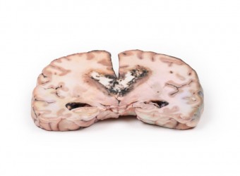
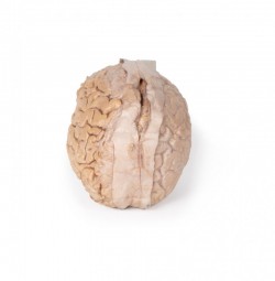
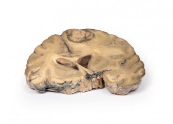
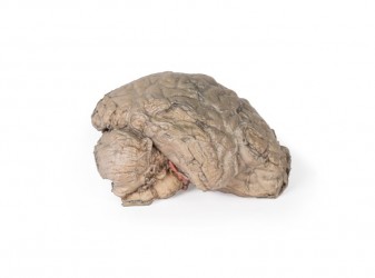
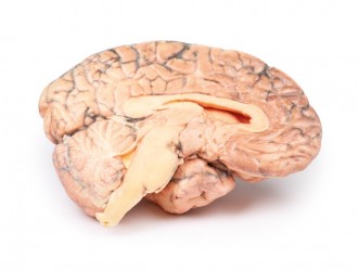
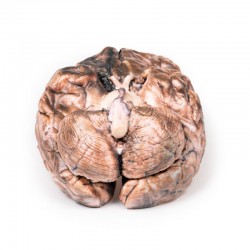
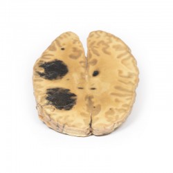
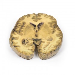
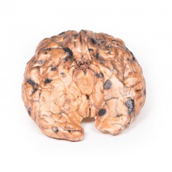
Astrocytoma
Quotation: +48 605 999 769
Delivery time and shippoing cost: please contact us:
+48 605 999 769, kontakt@openmedis.pl
Inquiry Quotation: +48 605 999 769 Download a PDF file Add to quotation - wish list
Product description: Astrocytoma

Clinical History
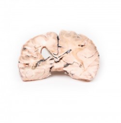
A 73-year-old female was admitted with new left-sided hemiplegia. On further questioning she revealed a 3-month history of headaches, nausea and deteriorating balance. CT brain revealed an inoperable brain tumour. She died 1 week after being admitted.
Pathology
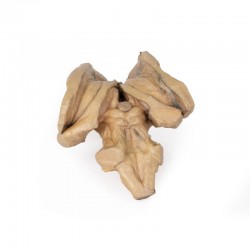
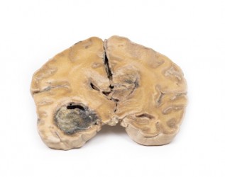
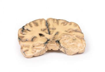
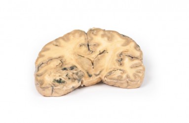
This brain specimen is a coronal section. In the right temporal lobe, a poorly demarcated tumour is present. There is enlargement of the hemispheres and flattening of the gyral pattern. From the posterior aspect of the specimen subfalcine herniation* is appreciated and the tumour appear less well differentiated with haemorrhagic and necrotic foci. Histology of this tumour showed an astrocytoma, Grade III/IV. *In subfalcine (or cingulate) herniation, the most common type of brain herniation, the innermost part of the frontal lobe is pushed under part of the falx cerebri, between the two hemispheres of the brain.
Further Information
Gliomas are the second most common cancer of the central nervous system after meningiomas. The term “glioma” refers to tumours that are histologically similar to normal glial cells i.e. astrocytes, oligodendrocytes and ependymal cells. They arise from a progenitor cell that differentiates down one of the cell lines. Astrocytomas develop from the astrocyte lineage of glial cells. Tumours are staged according to histological differentiation and range from diffuse astrocytoma (Grade II/IV) to anaplastic astrocytoma (Grade III/IV) to glioblastoma (Grade IV). Histological features include the prominent eosinophilic cytoplasm in some astrocytic tumour cells (gemistocytes) as well as a fibrillary background.
Astrocytomas occur most commonly between the fourth and sixth decades of life. Tumours usually occur in the cerebral hemispheres but may also occur in the cerebellum, brainstem or spinal cord. They most commonly present with seizures, headaches, nausea and focal neurological deficits depending on area involved. Without treatment Grade III median survival is 18 months. Treatment includes surgical resection, radiotherapy, chemotherapy or a combination thereof, depending on the clinical context.
Inquiry
Related products

Information
Purchase regulations
Privacy policy
Quality policy
About us
Cooperation
Registration
Login
Categories
Anatomical Models Anatomical Charts 3D anatomy models Anatomical Table 3D Medical simulators Veterinary simulators Custom tools for patient education Medical Equipment
Contact us
OpenMedis Sp.z o.o
ul. Kolejowa 2,
30-805 Kraków
NIP: PL6793229260