Send to quote
Cart (0)
Quality Certyficate street: Kolejowa 2, 30-805 Cracow
OpenMedis - Anatomial Models

Pituitary Adenoma

Product code: AM02708

Product description: Pituitary Adenoma

Clinical History

A 29-year old male presented with a 22 month history of headaches and blurred vision. Examination revealed a bi-temporal hemianopia and a left VIth nerve palsy. Skull X-ray showed erosion of most of the sphenoid body with some dorsum sellae and anterior clinoid process intact. Carotid angiography showed upward and lateral displacement of the anterior and middle cerebral arteries. Pneumoencephalography (a common imaging procedure used until the 1970s in which CSF is drained and replaced by air, oxygen or helium that acted as a contrast medium in X-ray examinations) showed upward displacement of the lateral and third ventricles from below. A craniotomy was performed but the patient died immediately afterwards

Pathology

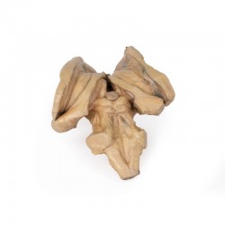
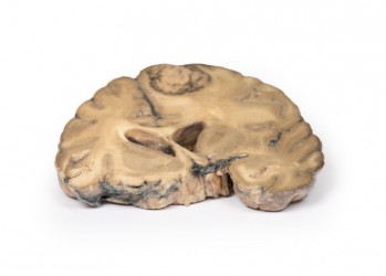
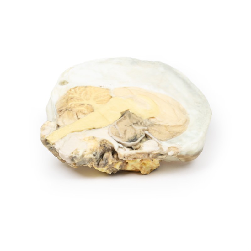
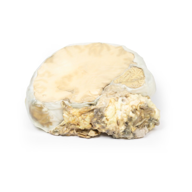
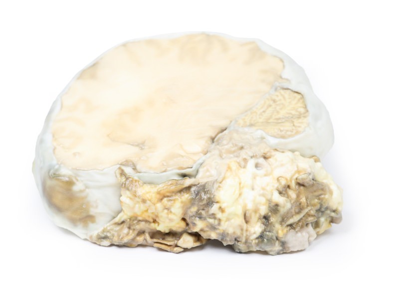
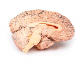
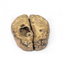
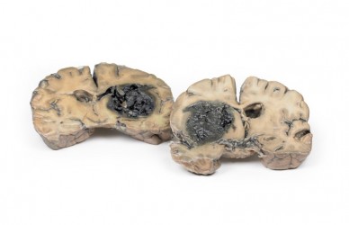
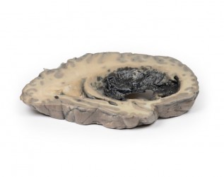
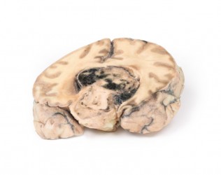
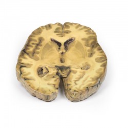
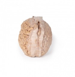
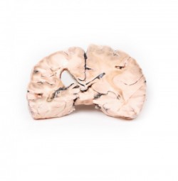
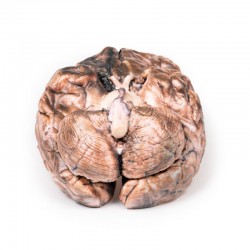
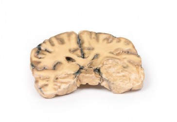
The brain specimen is sliced in the sagittal plane to the right of the falx cerebri, which remains in-situ. The pituitary gland has been completely replaced by a round tumour 4cm in maximum diameter. The tumour cut surface is pale brown and homogenous (except for an area of haemorrhage superiorly, likely caused by surgical trauma). The tumour has resulted in upward displacement of the midbrain. Tumour erosion has destroyed the sphenoid bone; thus, the sella turcica is enlarged (arrow). The optic chiasma is compressed by the tumour. Histologically, this tumour was a chromophobe adenoma arising from the anterior pituitary.

Further Information 
This specimen is from an old case and the investigations used would now be considered antiquated. Modern investigation would include an initial brain CT followed by an MRI of the brain to further visualise the pituitary lesion prior to any surgical intervention. Pituitary adenomas are the most common pituitary tumour and are most commonly found in adults with peak incidence between 35-60 years. Primary carcinoma of the pituitary is very rare, and the pituitary is an uncommon site for metastases. Clinical manifestations of pituitary adenomas are related to local mass effect and tumour function. Local effects include increasing intracranial pressure (headache, nausea and vomiting), sellar expansion, bony erosion and compression of decussating nerve fibres in the optic chiasma, causing bitemporal hemianopia. Pituitary adenomas can be functioning (i.e. associated with hormone excess) or non-functioning (i.e. without clinical symptoms of hormone excess). About 75% of adenomas are functional: usually secreting prolactin, growth hormone or ACTH. Secretion of TSH, LH and FSH from pituitary adenomas is very rare. Some adenomas can secrete two hormones: growth hormone and prolactin being the most common combination. Non-functional pituitary adenomas come to clinical attention later than those associated with endocrine abnormalities, and they may lead to hypopituitarism due to compression atrophy of the surrounding normal gland.
 

Inquiry

Your chosen product: Pituitary Adenoma
Name and surname:
Phone number:
E-mail adress:
Message:
I consent to the processing of my personal data within the meaning of the Act of 10 May 2018 on the Protection of Personal Data and the Act of 16 July 2004 Telecommunications Law for marketing purposes by OpenMedis and OpenMedis Sp. z o.o and I declare that the provision of my personal data is voluntary and that I have been informed about the right to request access to my personal data, change and delete it.

Related products

Openmedis - recommended categories