


Home / 3D anatomy models / 3D anatomical models of the brain / Berry Aneurism of Basilar Artery
Berry Aneurism of Basilar Artery
Berry Aneurism of Basilar Artery
 Download a PDF file Add to quotation - wish list
Product description: Berry Aneurism of Basilar Artery

Clinical History
A 37-year-old patient presented to hospital after falling and striking his head, with subsequent symptoms of headache, vomiting and disorientation. CT scan showed dilatation of the lateral ventricles associated with a large mass projecting into the third ventricle posteriorly. One week later a shunt was performed for hydrocephalus. An angiogram revealed a partially thrombosed aneurysm, measuring 1 x 1 cm, arising from the basilar artery. At 3-months post-op the shunt was revised due to obstruction, with repeat cerebral angiogram revealing interval enlargement of the aneurysm. Attempted ligation of the aneurysm was unsuccessful. The patient remained unconscious despite several attempts to revise the shunt and he died.
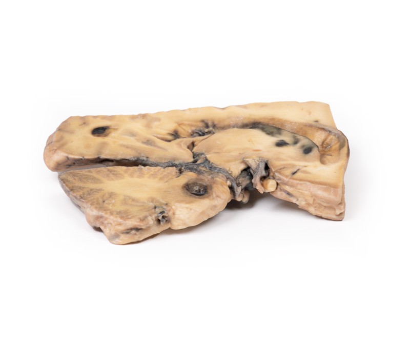
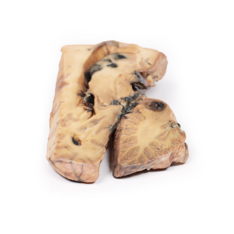
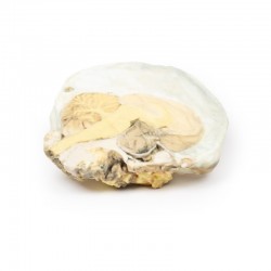
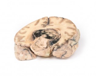
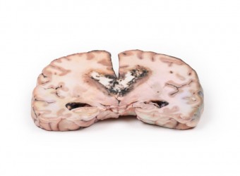
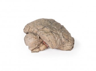
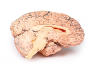
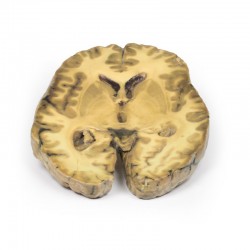
Pathology
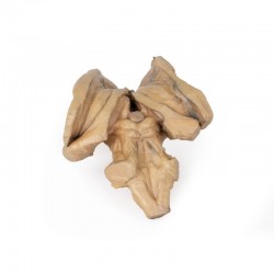
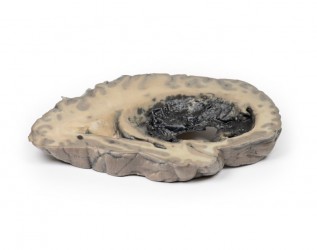
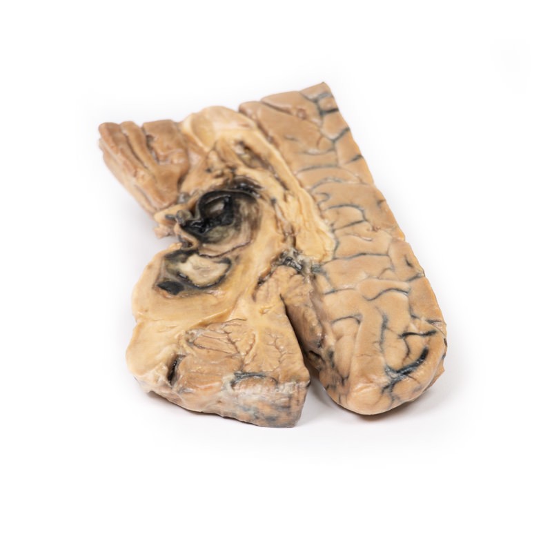
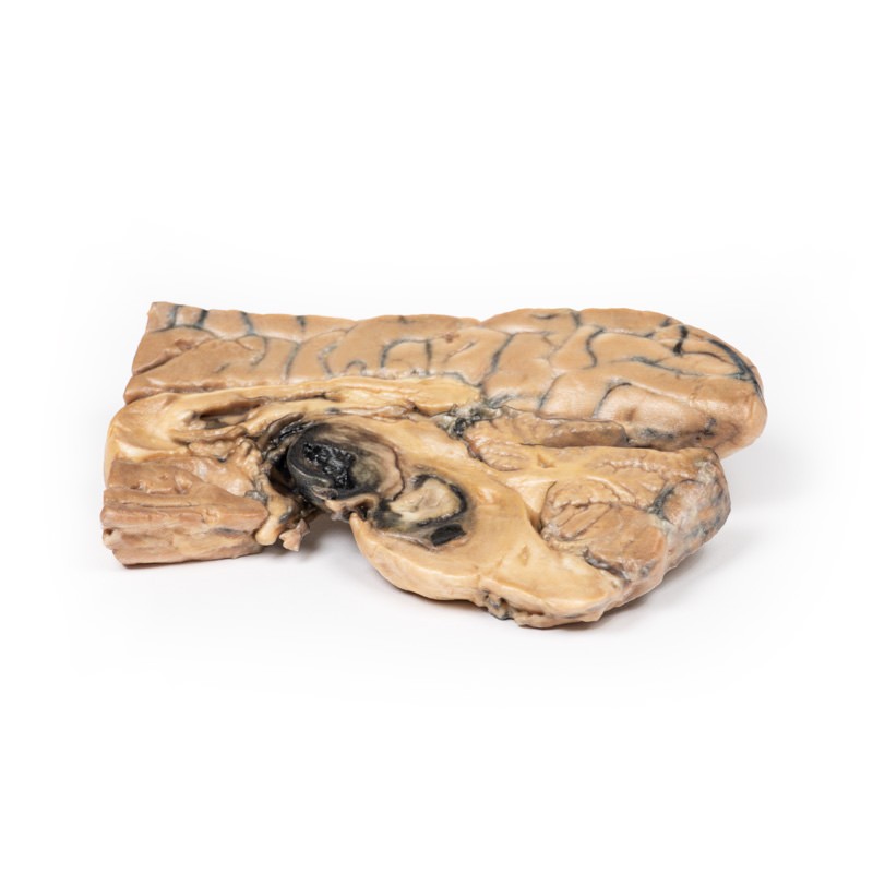
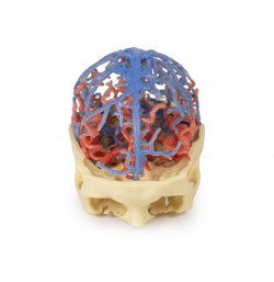
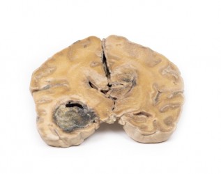
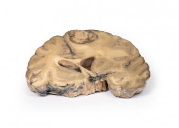
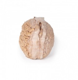
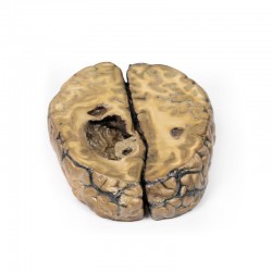
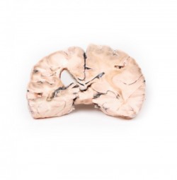
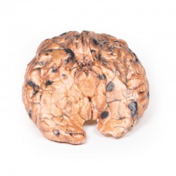
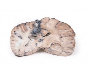
This brain has been sliced in the mid-sagittal plane. It comprises a whole hemi-section of the brain about 1cm thick. On the medial surface a large darkly-coloured ovoid berry aneurysm measuring 5 x 2 cm in diameter, arising from the basilar artery is clearly visible. It has eroded up into the midbrain, compressing the third ventricle from below, and inferiorly into the substance of the pons. The wall of the aneurysm appears intact although blood clot is seen in the third ventricle and appears to be leaking through the lateral wall of that ventricle. The aneurysm is filled with a laminated thrombus. A small area of mucoid degeneration measuring 0.4 cm in diameter is seen posterior to the aneurysm within the pons. Examination of the lateral aspect of the sagittal section shows dilatation of the lateral ventricle, blood staining of the ventricular wall and patchy haemorrhagic infarction of the caudate nucleus. There was some discolouration of the meninges overlying the tip of the left temporal lobe and the cerebellum (not included in 3D print), consistent with sub-arachnoid haemorrhage.
Further Information
Prevalence of aneurysms is approximately 3.2% in the population, while rupture is much less common, occurring only 7.9 per 100,000 person-years. A minority of intracranial aneurysms arise from the posterior circulation, and are mostly situated at junctional points about the basilar, vertebral and cerebellar arteries. Symptoms are either secondary to subarachnoid haemorrhage or a mass effect with associated compression of the adjacent brain parenchyma and cranial nerves. Rupture causes complications due to bleeding and raised intracranial pressure. Hydrocephalus, re-bleed and vasospasm may also occur. Management is via surgical means; in recent years, novel therapies include endovascular intervention with coils and subsequent monitoring.