


Home / Anatomical Models / Upper extremities models / Hand anatomical model, 7 parts
Hand anatomical model, 7 parts

Hand anatomical model, 7 parts
 Download a PDF file Add to quotation - wish list
Product description: Hand anatomical model, 7 parts

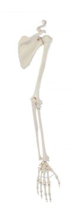
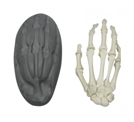
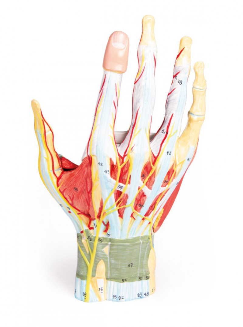
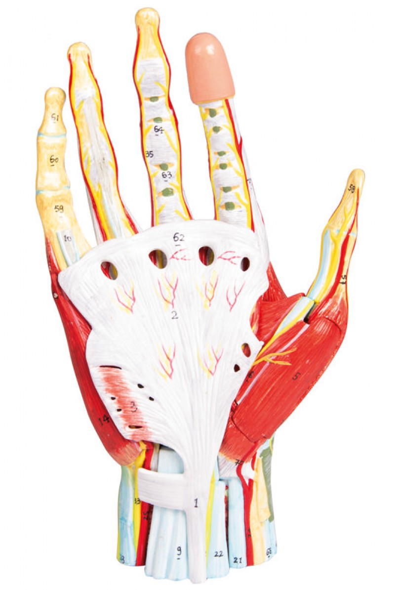
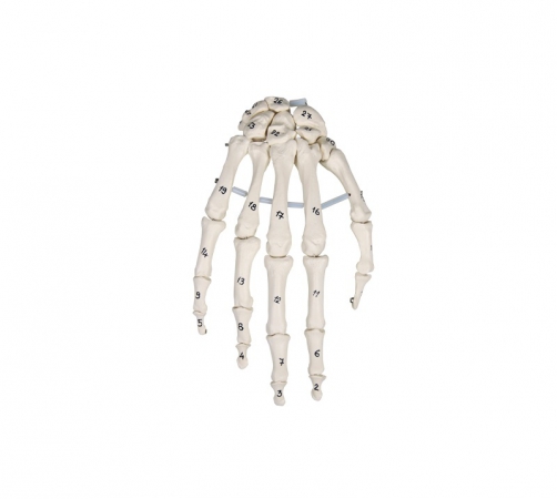
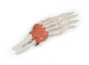
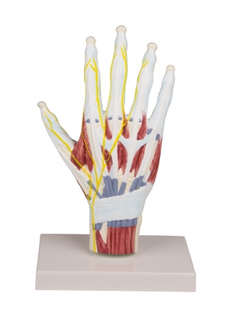
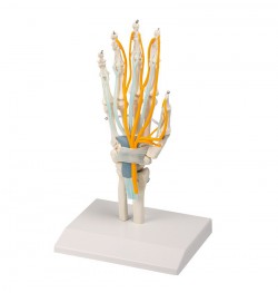
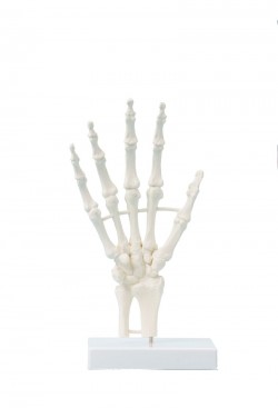
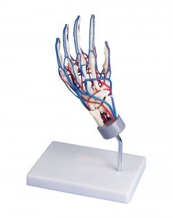
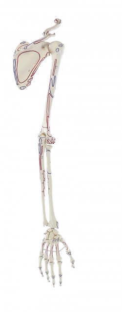
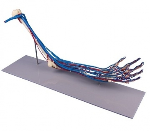
The anatomical model of the right human hand is an ideal teaching aid for medical students who want to explore the details of the anatomical structure of the hand. The model is made entirely of artificial material, thanks to which it can be used in ordinary rooms and does not wear out over time, ensuring the same educational conditions all the time. The use of varied colors to paint individual elements adds didactic value to the presented model. The hand model facilitates the study of the topography of the anatomical structures of the hand and the mutual relations between them. Thanks to the use of discreetly hidden magnets, you can easily unfold and fold the model by analyzing the anatomical structure of the hand. The hand model can be disassembled into 7 separate elements. It presents important anatomical structures running within the hand: muscles of the withers, glomerulus, interosseous, ascending muscles, tendons, nerves, blood vessels. In addition, the model has the numbering of individual elements.
.jpg)
Advantages of the human hand anatomical model:
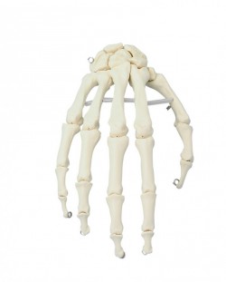
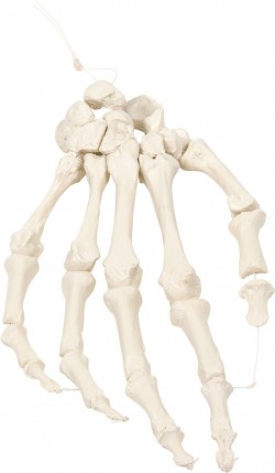
- The hand model can be disassembled into 7 parts
- Diversified colors, thanks to which the individual structures are perfectly exposed
- Learning the topography of anatomical structures of the hand and mutual relations between them
- Structure numbering
Hand model application
- Anatomy lab equipment
- Learning the anatomy of the human hand
- The anatomical model can be used to demonstrate the structure of the human hand - to patients in office conditions
- As a teaching aid during postgraduate training for doctors, physiotherapists, osteopaths and other medical professions.
Specification:
- Size: 11 x 13 x 33 cm
- Weight: 0.5 kg