


Home / 3D anatomy models / 3D anatomical models of upper limb / Forearm and Hand - superficial and deep dissection
Forearm and Hand - superficial and deep dissection


Forearm and Hand - superficial and deep dissection
 Download a PDF file Add to quotation - wish list
Product description: Forearm and Hand - superficial and deep dissection

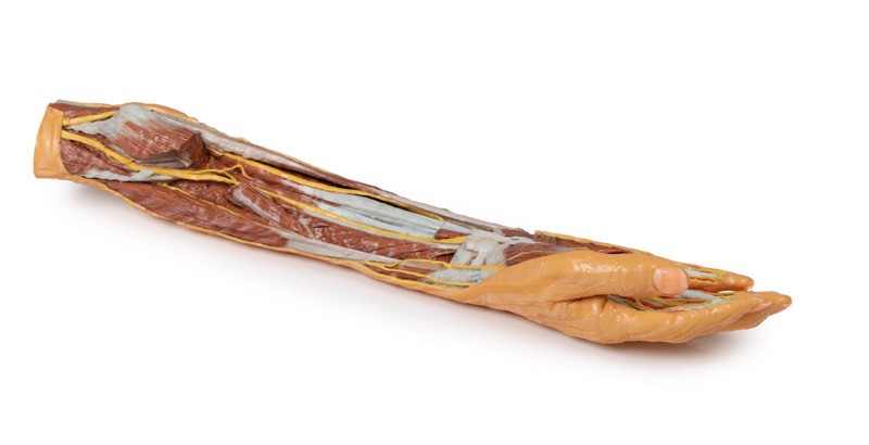
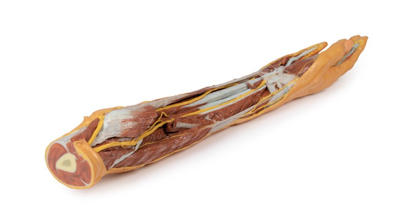
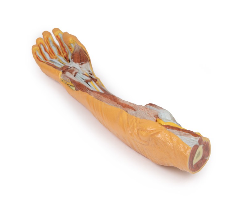
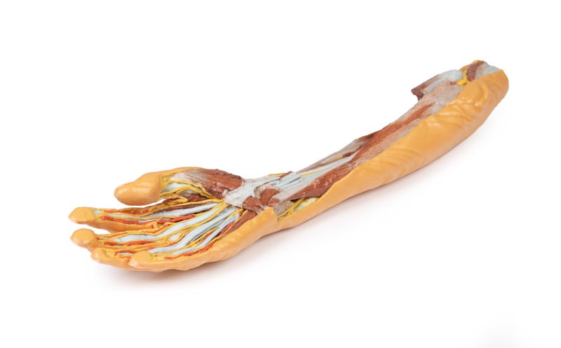
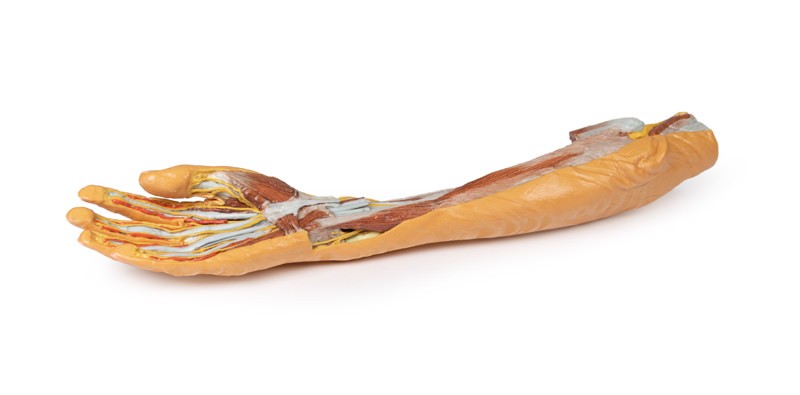
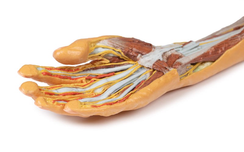
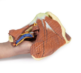
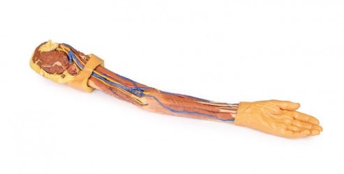
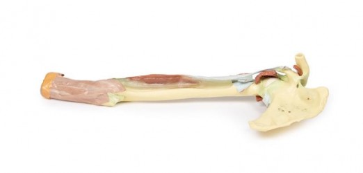
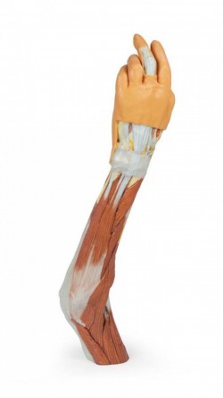
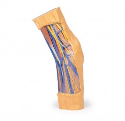
This 3D printed specimen preserves a mixed superficial and deep dissection of the anterior aspect of a right distal arm, forearm and hand. Proximally and medially the ulnar nerve passes through the cubital tunnel before passing deep to the flexor carpi ulnaris. The cubital fossa has been opened by removing most of the flexor musculature to demonstrate the insertion of the biceps brachii and brachialis muscles, and the course of the median, ulnar and superficial branch of the radial nerve in the anterior compartment of the forearm. Several muscular branches of the median nerve are visible entering the prontator teres and flexor digitorum profundus muscles. The insertion of the pronator teres has been retained, lying between the partially exposed supinator and the flexor pollicis longus muscles. Near the wrist, the tendons of the flexor digitorum superficialis have been retained to demonstrate their passage through the carpal tunnel alongside the tendons of the flexor digitorum profundus, flexor pollicis longus, and the median nerve. Deep to all these muscles, part of the pronator quadratus muscle is visible. The flexor retinaculum itself is covered by the palmaris longu tendon, base of the palmar aponeurosis, and a prominent palmaris brevis muscle. Laterally, the superficial branch of the radial nerve crosses the extensor pollicis brevis and abductor pollics longus tendons to enter the roof of the anatomical snuffbox. The hand itself is superficially dissected, with only the skin, subcutaneous fascia and palmar aponeurosis removed between the thenar and hypothenar eminences. The flexor digitorum superficialis and profundus tendons extend through the palm, surrounded by lumbricals and terminal branches of the median and ulnar nerves into the common and proper digital nerves. Most of the palmar arterial structures have been removed to improve the visibility of the fine structures in the hand, however a single common palmar digital artery has been retained as have the proper palmar digital arteries along the sides of the digits.
Inquiry
Related products

