


Home / 3D anatomy models / 3D anatomical models of upper limb / Model of upper limb ? bones, ligaments
Model of upper limb ? bones, ligaments

Model of upper limb ? bones, ligaments
 Download a PDF file Add to quotation - wish list
Product description: Model of upper limb ? bones, ligaments

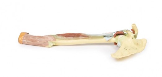
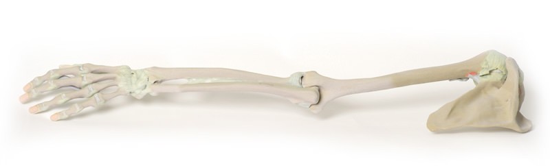
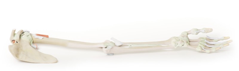
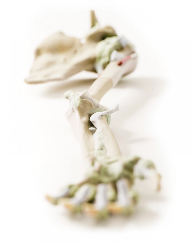
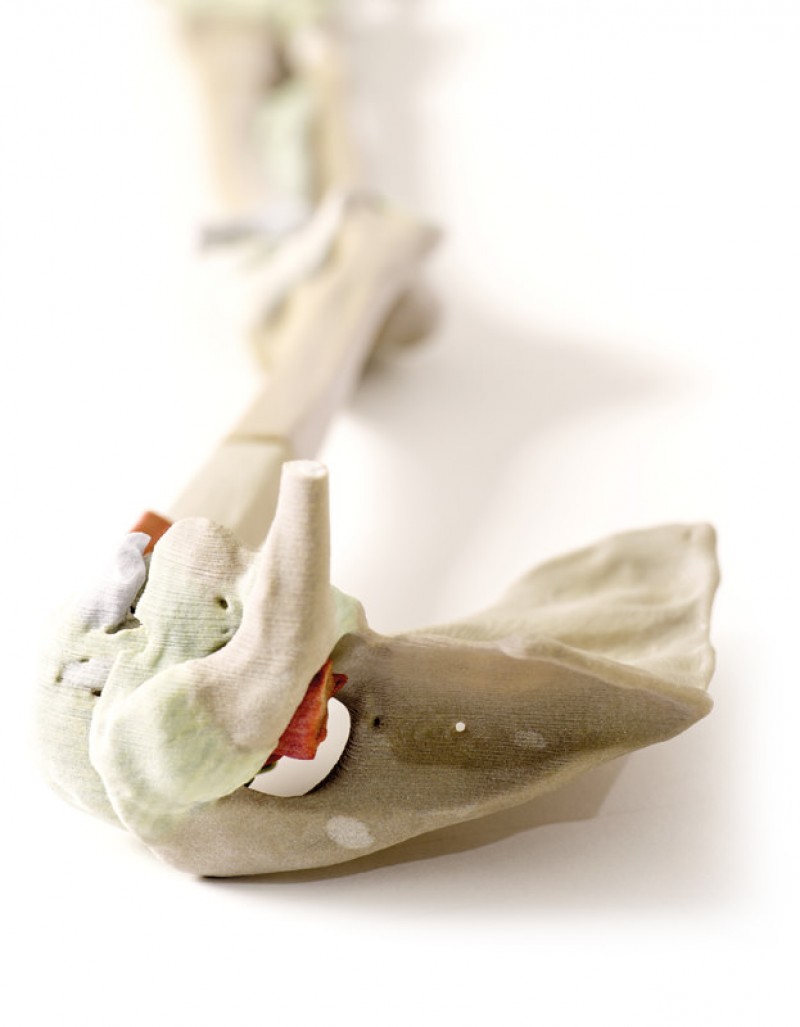
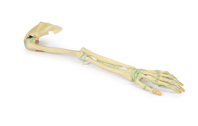
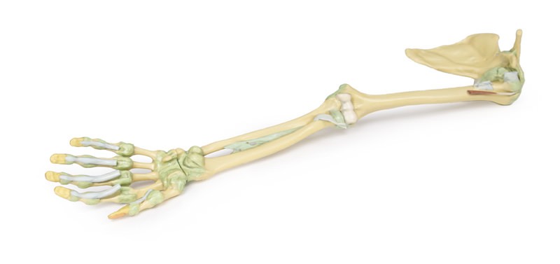
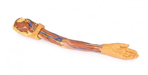
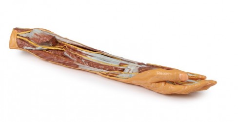
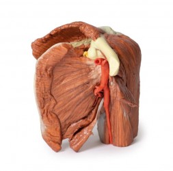
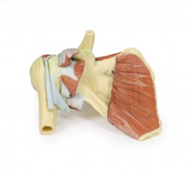
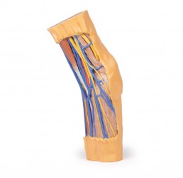
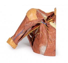
This 3D printed specimen presents the entire upper limb skeleton and ligaments from the pectoral girdle to the hand. In the pectoral girdle, the ligaments spanning the clavicle and scapula (acromioclavicular, coracoclavicular, coracoacromial) as well as the superior transverse scapular ligament spanning the suprascapular notch, are visible. A small portion of the supraspinatus muscle belly and tendon are preserved to demonstrate the passage of the muscle deep to the coracoacromial ligament, which is a very clinically relevant area of anatomy. The tendon of the subscapularis muscle has been reflected slightly to expose the anterior aspect of the glenohumeral joint capsule, and the tendon of the long head of triceps brachii, teres major, and latissimus dorsi are preserved surrounding the capsule and proximal humerus. The tendon of the long head of biceps brachii is visible within the intertubercular groove, and exposed within the superior glenohumeral joint capsule as it approaches the supraglenoid tubercle.
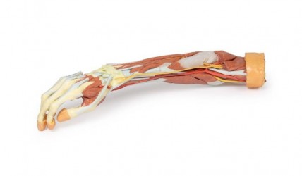
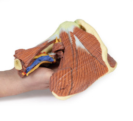
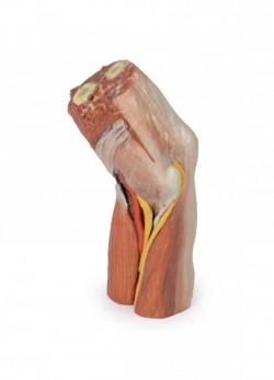
The capsule of the elbow joint has been dissected to expose the articular surfaces of the distal humerus, proximal radius and proximal ulna. Both the ulnar and radial collateral ligaments are preserved, as is the annular ligament of the radius. Just distal to the joint capsule, the tendinous insertion of the biceps brachii is preserved as it inserts into the dorsal aspect of radial tuberosity.
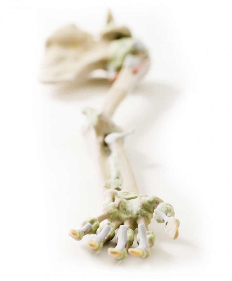
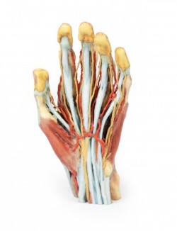
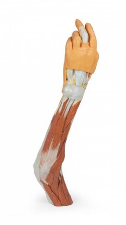
Distal to the interosseous membrane, the palmar and dorsal ligaments of the wrist joint are preserved (including the radial and ulnar collateral ligaments, palmar and dorsal radiocarpal and ulnocarpal ligaments, pisohamate, pisometacarpal, radiate capitate, palmar and dorsal carpometacarpal ligaments). In the hand, the metacarpophalangeal and interphalangeal joint capsules with collateral ligaments are preserved for all digits, including the palmar ligaments (volar plates); the capsules are open dorsally to appreciate the articulations between elements. The terminal portions of the flexor digitorum superficialis and profundus tendons are retained to show their insertions into the bases of the intermediate and distal phalanges, as is the flexor pollicis longus tendon inserting to the base of the terminal phalanx of the first digit.
Inquiry
Related products
