Anatomical Models / Pelvis models
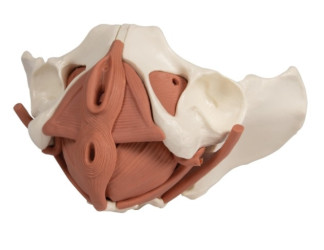
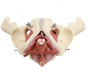
A life-size model of the pelvic floor muscles. Individual muscles can be easily removed. The model also shows the bone anatomy of the female pelvis. The model presents the following structures:
Obturatorius ...
This pelvis is eminently suited to explanation of the female pelvic floor. The model consists of 2 hip bones, sacrum with coccyx and the pelvic floor. The pelvic floor is made of flexible synthetic material on which ...
This pelvis is eminently suited to explanation of the male pelvic floor. The model consists of 2 hip bones, sacrum with coccyx and the pelvic floor. The pelvic floor is made of flexible synthetic material on which the ...
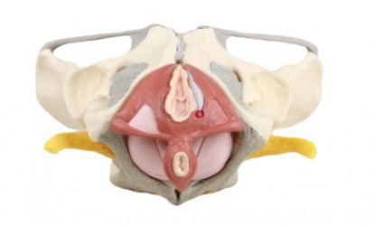
This pelvis is eminently suited to explanation of the female pelvic floor. The model consists of 2 hip bones, sacrum with coccyx and the pelvic floor. The pelvic floor consists of 4 components and is made of flexible ...
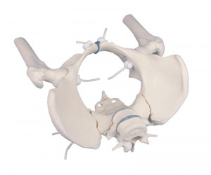
Female pelvis model with ligaments. This model is not dissectible and shows the position and function of the ligaments in the female pelvis. Life size. ...
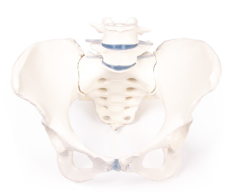
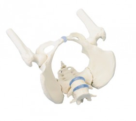
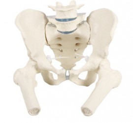
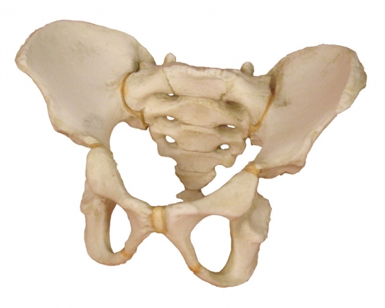
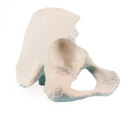
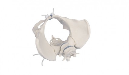
Natural casting of an adult female pelvis.
The sacrum is removable and the movements in the iliosacral joint can be demonstrated.
The vertebrae are mobile mounted and the femoral stumps are ...
Natural casting of an adult male pelvis.
The sacrum is removable and the movementsin the iliosacral joint can bedemonstrated.
The vertebrae are mobilemounted and the femoral stumps ...
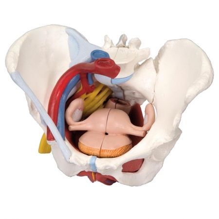
This life size six part model of a female pelvis represents detailed information about the topography of bones, ligaments, vessels, nerves, pelvic floor muscles and female genital organs. It presents the whole pelvic ...
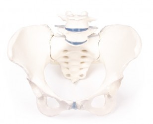
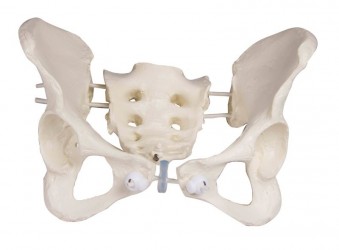
Natural casting of an adult female pelvis. wings of ilium, sacrum and flexibly mounted L5 and L4.
The sacrum is removable and the movements in the iliosacral joint can be demonstrated.
...
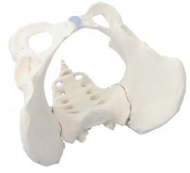
Natural casting of an adult male pelvis.
Wings of ilium, sacrum and flexiblymounted L1 and L2.
The sacrum isremovable and the movements in theiliosacral joint can be demonstrated.
...
A Life-size male pelvis model with sacral bone showing bone anatomy. The Examples of structures shown in the model are, for example, anterior superior iliac spines, anterior lower bidra spines, sciatic tumors, ischial ...
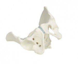
Actual cast of a real human bony child pelvis.
This model is particularly suitable for explanation of the pelvis development during growth.
The one-part model is not movable.
...
The model shows the female genitalia, bladder with ureter and anus.
Extended anatomy app:
Learning is now even easier and more efficient with the new Augmented Anatomy app in conjunction with this high-quality ...
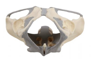
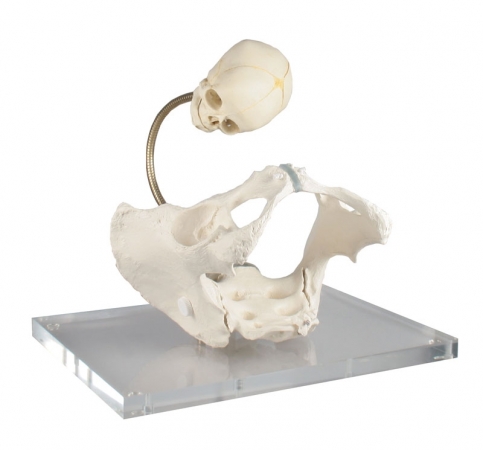
With this model birth canal and the passage of the fetal head through the female pelvis during labour can be clearly demonstrated.
The pelvic skeleton consists of flexible hip bones with movably ...
A life-size cast of the female pelvis. It has the ability to disassemble the sacrum and demonstrate mobility in the sacroiliac joint. The model also has two lower lumbar vertebrae. Fragments of the femurs move ...
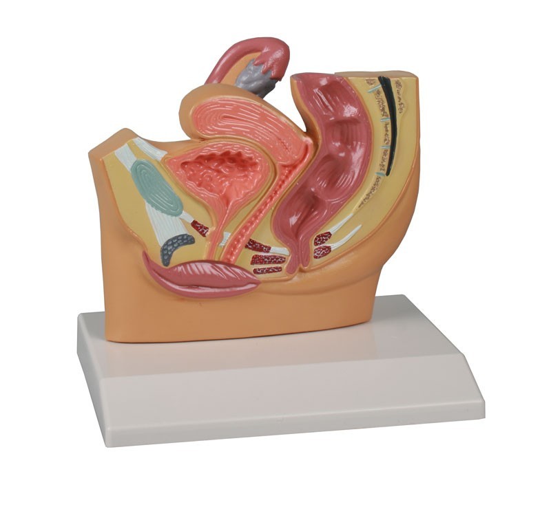
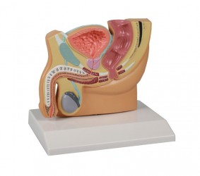
The male pelvis model, reduced twice, shows:
male reproductive system
bladder
prostate
urethra
rectum
...
Excellent casting of the bones of an adult male. ...
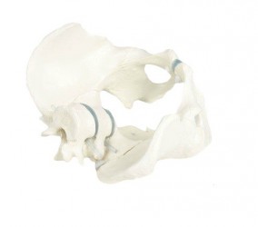
A natural cast of an adult woman's pelvis. The sacrum is removable, movement in the sacroiliac joint is natural. ...
Natural casting of an adult female pelvis. The sacrum is removable and the movements in the iliosacral joint can be demonstrated. ...
The model shows a natural cast of an adult woman's pelvis. The sacrum can be removed and movements in the sacroiliac joint can be demonstrated. ...
1
Anatomical Models - Pelvis models
Anatomical models of the human pelvis are highly useful teaching aids for all those who want to deepen their knowledge about the bone structure and organs located in the human pelvic area. Using different colors to paint individual elements makes it easier to learn the topography of the anatomical structures of the human pelvis and the mutual relations between them. Thanks to the use of the highest quality materials and advanced production methods, human pelvic models are characterized by high durability and precision. Many bone models are natural casts of human pelvises. Our assortment includes both bone models of the pelvis as well as cross-sections and models presenting organs located in the human pelvis. Depending on the type, anatomical models of the pelvis present superficial structures and the bony anatomical structure of the pelvis, while others present the muscular structure of the pelvic floor, the course of blood vessels and nerves in this complex region. Some anatomical models can be broken down into individual elements, which makes learning anatomy much more attractive and easier. Some models of the human pelvis include models of internal organs, thanks to which you can easily learn about the topography and mutual relations between individual structures.
Our offer includes, among others: the following anatomical models of the pelvis:
- Pelvic floor muscle model, 12 parts
- Model of the female pelvic floor muscles
- Model of the male pelvic floor muscles
- Model of the pelvis with nerves and ligaments
- Cross-sectional model of the male pelvis
- Cross-sectional model of the female pelvis
- Pelvic models with removable and disassembled organs
- Bone models of the pelvis
- Pelvis models with lower lumbar vertebrae
- Pelvis models with proximal femur fragments
- Pelvic model for childbirth demonstration
- Flexible pelvic models showing mobility
The human pelvis model is ideal for learning anatomy at home and during seminars conducted at universities and medical schools. Based on radiological data, a series of models faithfully reflecting the human body were created. These advanced teaching aids are the equipment of anatomy laboratories of many Medical Universities in Poland. This is a modern solution used in many countries around the world.
Advantages of anatomical models of the human pelvis:
- Accuracy of workmanship (hand-finished)
- Durability (made of the highest quality materials)
- Learning the anatomy of the normal human pelvis
- Learning the topography of anatomical structures located in the human pelvis and the mutual relations between them
Application of human pelvis model:
- Equipment of the anatomy laboratory
- Learning the anatomy of the human pelvic region
- The anatomical model can be used to demonstrate the structure of the human pelvis - to patients in an office setting
- As a teaching aid during postgraduate training for doctors, physiotherapists, osteopaths and other medical professions
The anatomical models of the pelvis offered by our store are made of high-quality materials, detailed and durable. Thanks to their realistic structure, they enable effective learning and construction demonstration. Our anatomical models are aimed at students, doctors and medical specialists and are a perfect complement to all educational and research needs.
We also recommend
skull models and
spine models, which, combined with anatomical pelvis models, will create an ideal set for learning anatomy.
If you have any questions or if the pelvis model you need is not available in the store, please contact us via e-mail or telephone. We are sure that together we will be able to provide the necessary materials for learning and improving skills. We can produce custom models with individual specifications.


 See our profile on Facebook
 Check our profile on Instagram
 Download a PDF file
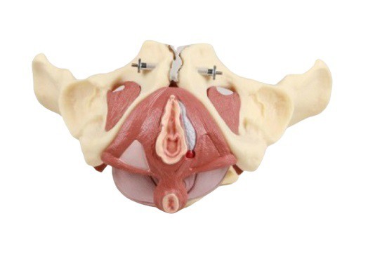


