Quality Certyficate
street: Kolejowa 2, 30-805 Cracow


Home / 3D anatomy models / 3D anatomical models of the brain
3D anatomy models
Anatomical Models
Custom tools for patient education
Veterinary simulators
Anatomical Charts
Anatomical Table 3D
Medical simulators
Medical Equipment
Type:
 See our profile on Facebook
 Check our profile on Instagram
 Download a PDF file
3D anatomy models / 3D anatomical models of the brain
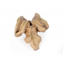
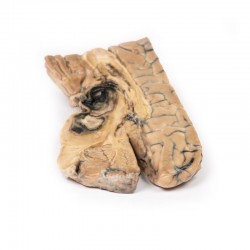
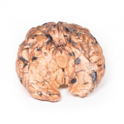
This 3D brain stem anatomical model preserves the several deep cerebral and diencephalic structures through to the proximal medulla oblongata and compliment the other isolated brainstem (BRW10) in our series. ...

This 3D model provides a view of the isolated brainstem anatomy from the midbrain to the medulla oblongata, and compliments the other diencephalon/brainstem 3D model (BR 10) in our series. Rostrally, the 3D model has ...
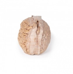
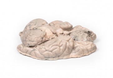
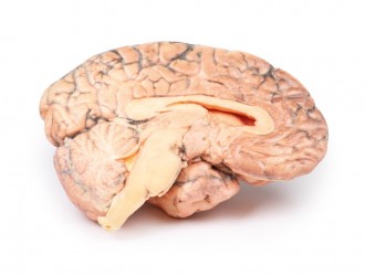
This 3D model provides a unique perspective on the anatomy of the cerebrum relative to the meninges. The cerebrum has been separated from the brainstem and cerebellum, with only parts of the midbrain and cerebral ...
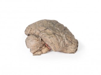
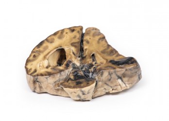
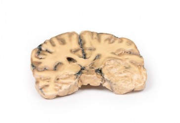
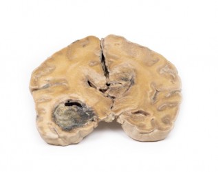
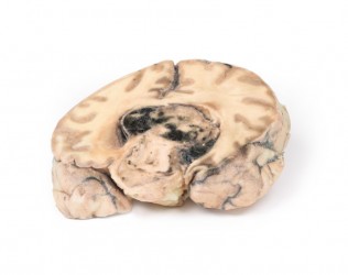
This 3D model is a midsagittal hemisection through a whole brain, preserving the right side anatomy and deep brain structures and spaces visible in the midline. In lateral view, the right cerebral and cerebellar ...
Clinical History
A 37-year-old patient presented to hospital after falling and striking his head, with subsequent symptoms of headache, vomiting and disorientation. CT scan showed dilatation of the lateral ventricles ...
Clinical History
A woman of 56-years was admitted following 2 episodes of severe headache with loss of consciousness. Clinical examination revealed systemic hypertension with cardiac enlargement, and a right ...
Clinical History
The patient was a female aged 24 years, who presented 18 months before death with an abnormal electroencephalogram (EEG) after a single epileptic fit. Six months later she complained of blurred vision ...
Clinical History
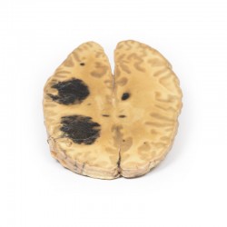
A 68-year-old female presented with recent onset of seizures and was diagnosed with epilepsy. Collateral history revealed a gradual change in the patient’s personality. She subsequently died several ...
Clinical History
The patient was a 51-year old woman who had a cerebrovascular accident resulting in a left hemiplegia 2 years prior to death. At necropsy, she had severe generalized atherosclerosis and an old ...

Clinical History
A 29-year old male presented with a 22 month history of headaches and blurred vision. Examination revealed a bi-temporal hemianopia and a left VIth nerve palsy. Skull X-ray showed erosion of most of ...
Clinical History
A 56-year old male underwent a total gastrectomy and splenectomy for gastric adenocarcinoma. Over a period of two months he developed a progressively unsteady gait, increasing weakness of his left hand ...
Clinical History
A 50-year-old alcoholic was admitted with a 2-week history of weakness and shortness of breath. At the onset of the illness he reported a productive cough, chest pain and blood-stained sputum. ...
Clinical History
A 22-year-old male presented with a 2-week history of generalised malaise, weight loss and bruised skin without any trauma. He recently developed 5 days of productive cough and fevers. He was ...
Clinical History
This patient died at the age of 58 years from post-operative complications following transurethral resection of the prostate. At the ages of 28 and 35, he had suffered two episodes of transient ...
Clinical History
A 56-year-old woman with 6 months of intermittent headache and vomiting was admitted to hospital comatose after a grand mal seizure, and failed to regain ...
Clinical History
The patient was an 80-year old man who suddenly lost consciousness. On examination there was a right gaze palsy, a left hemiplegia and right hemiparesis.
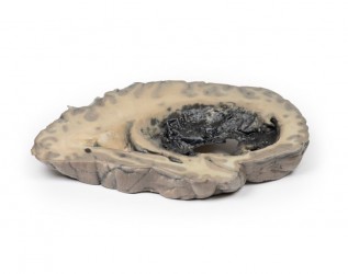
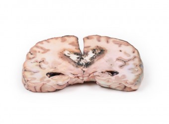
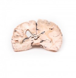
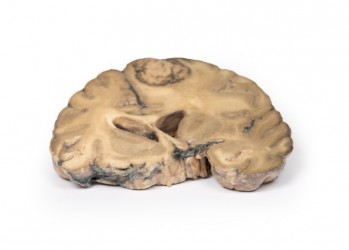
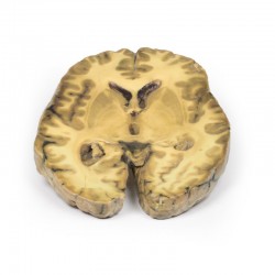
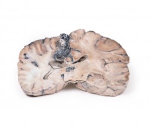
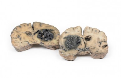
Pathology
The specimens are coronal ...
Clinical History
Five days before admission this 38-year old female experienced the sudden onset of pain behind the right eye, associated with a slow development of weakness of the left leg. Examination disclosed ...
Clinical History
A 73-year-old female was admitted with new left-sided hemiplegia. On further questioning she revealed a 3-month history of headaches, nausea and deteriorating balance. CT brain revealed an ...
Clinical History
Over a 3-year period, a 57-year-old woman had intermittent frontal headache and memory disturbance with progression to psychiatric disturbance, and ultimately vomiting and meningeal signs. ...
Clinical History
A 56-year old male presented with a generalised seizure. He remained unconscious after this seizure and later died. A collateral history revealed 6 months of progressive confusion, short-term ...
Clinical History
A 62-year-old woman presented with disorientation to time, place and person. Physical examination revealed no localised neurological signs. Radiological investigations revealed a space occupying ...
Clinical History
In the 1970s, a 31-year-old woman presented with severe headache and diplopia on a background of having a pigmented skin lesion (diagnosed as an invasive skin melanoma) removed from her neck 8 ...
Clinical History (pre access to CT and MRI imaging)
This 51-year old woman had surgery for breast carcinoma 2 years before presentation. Her main complaint was left-sided ataxia for the 2 weeks prior, and this had ...
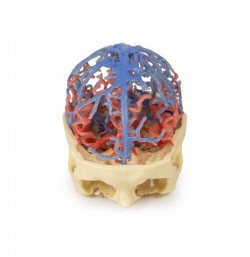
: This 3D print integrates segmented angiographic data of both the cranial arterial and venous circulation into a single model. Further description of the visible structures can be found under the ‘Circle of ...
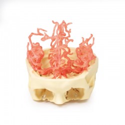
This 3D print presents the same dataset that underlies our circle of Willis and cranial arterial circulation 3D prints and is derived from careful segmentation of angiographic data. Here, the dural venous sinus network ...
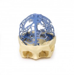
This 3D print presents an expanded version of the same dataset that underlies our circle of Willis 3D print derived from careful segmentation of angiographic data. Like our circle of Willis print, this model ...
1
3D anatomy models - 3D anatomical models of the brain
3D anatomical models of the brain as a teaching tool:
3D anatomical models of the brain are excellent educational tools, presenting the complex structure of this organ in three dimensions. Thanks to the precise reproduction of anatomical details, these models allow for a deeper understanding of the structure of the brain, its functions and complex neural connections. The brain can be divided into: cerebral cortex, white matter, basal ganglia, limbic system and many others. Each of these areas has its own role and functions. Taking into account the division, it should be noted that the human brain is a wealth of various anatomical structures and details, often running close to each other or intersecting each other. From a teaching point of view, it is important to carefully visualize these structures in order to understand their functions as well as the mutual relations between them. During the teaching process, precisely made anatomical models, faithfully presenting the human body, work great. These models allow you to visualize these structures and connections, making it easier to learn anatomy and neurology. In this product category, we present anatomical models of the brain created on the basis of CT radiological data and using modern 3D printing technology. OpenMedis also offers conventional anatomical models of the brain.
3D anatomical models of the brain - advantages:
It should be mentioned that 3D anatomical models of the brain are made using modern 3D printing technology, which allows for obtaining very accurate replicas. The artificial materials used to produce these models are durable and wear-resistant, which guarantees long-term use for teaching purposes. Thanks to the careful consideration of radiological data from tomographic (CT) imaging in the creation of these models, their accuracy in representing the real brain is impressive. These models show the brain in all its glory, including structures such as complex networks of neurons, blood vessels and functional areas. This far exceeds the capabilities of conventional models, which often simplify the anatomy of the brain. Another advantage of 3D anatomical models of the brain is the lack of the need for special storage conditions. They can be used in standard classrooms and medical offices. Moreover, they do not generate waste or the need for disposal, which makes them economical and environmentally friendly. There is no doubt that the colors of individual structures, tailored to educational purposes, facilitate learning anatomy by understanding the topography of individual structures and the mutual relations between them.
3D brain anatomical models - application:
3D anatomical brain models are an ideal tool for teaching anatomy and neuroscience at various educational levels. They serve as teaching aids for students of medicine, psychology, neurology and other fields related to biomedical sciences. Currently, it is a modern solution used at many universities around the world. 3D anatomical models of the human brain are perfect equipment for anatomical and biological laboratories and human anatomy museums. 3D brain anatomical models can also be used for diagnostic purposes, helping patients better understand their conditions.
Types of 3D brain anatomical models:
Our offer includes so-called anatomical models conventional and unconventional. In this product category, we would like to interest you in 3D anatomical models of the brain, which faithfully present its anatomical structure as on real dissection preparations. Various types of 3D brain anatomical models are available, covering various brain regions and functions. You can find models showing the cerebral cortex, hippocampus, cerebellum, amygdala and many other structures. Additionally, my offer includes models of various types of diseases and conditions related to brain tissue. Each of these models is precisely made and reflects the actual anatomy of the brain.
- The brain stem model and the left hemisphere infarction model are of great interest.
Based on radiological data, a series of models faithfully reflecting the human body were created. These advanced teaching aids are the equipment of anatomy laboratories of many Medical Universities in Poland. This is a modern solution used in many countries around the world.
How can we help?
We are an experienced manufacturer and distributor of 3D brain anatomical models on the market. Our models are highly appreciated by medical schools, educational institutions and medical institutions around the world. If you need advice on selecting appropriate 3D brain anatomical models for your institution or would like to learn more details about our products, please contact us.
We also recommend other anatomical models available in our offer, such as 3D head models and 3D torso models, which can complement the set of tools for learning the anatomy and physiology of the body.
