Quality Certyficate
street: Kolejowa 2, 30-805 Cracow


Home / 3D anatomy models / 3D eye area
3D anatomy models
Anatomical Models
Custom tools for patient education
Veterinary simulators
Anatomical Charts
Anatomical Table 3D
Medical simulators
Medical Equipment
Type:
 See our profile on Facebook
 Check our profile on Instagram
 Download a PDF file
3D anatomy models / 3D eye area

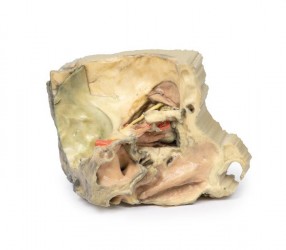
This 3D printed model captures a dissection in which the calvaria and cerebrum have been removed to expose the floors of the anterior and middle cranial fossae. The midbrain has been sectioned at the level of the ...

This 3D printed specimen shows the orbit from the lateral perspective when the bony lateral wall and part of the calvaria of the skull have been removed. The frontal and temporal lobes of the brain are exposed. In ...
This 3D print displays the orbital contents and its close relations as viewed from the medial perspective when the majority of the lateral wall of the nasal cavity and the intervening ethmoidal sinuses have been ...
1
3D anatomy models - 3D eye area
3D eye anatomical models as a teaching tool:
3D eye anatomical models are teaching aids that present precisely reconstructed eye structures, such as the cornea, lens, retina, eye muscles, blood vessels and nerves, in a three-dimensional form. Anatomically, the eye can be divided into various components, and these models allow for a thorough analysis of its structure and function. The elements that make up the anatomical model of the eye include: the cornea, iris, lens, vitreous body, retina, blood vessels, eye muscles and optic nerves. For teaching purposes, it is important to precisely present these structures in order to understand their role in the vision process and the relationship between them. In the process of learning eye anatomy, 3D eye anatomical models are an invaluable tool that faithfully reproduces the structures of the eye in a real way. In this product category, we present 3D eye anatomical models developed on the basis of radiological data and 3D printing technology. OpenMedis also offers traditional anatomical models of the eye.
3D eye anatomical models - advantages:
It is worth emphasizing that the 3D eye anatomical models are made entirely of durable and resistant artificial material. Thanks to this, they are extremely durable and do not wear out, ensuring uniform and fair teaching conditions for all students. An additional advantage of these models is their extremely precise manufacturing, possible thanks to the use of radiological data from computed tomography and 3D printing technology. These are high-fidelity models that accurately reflect the anatomy of the eye, including both external and internal structures. 3D anatomical models of the eye allow for a detailed examination of the cornea, lens, retina, and other important elements, which is a significant advantage over traditional models, which often present anatomy in a more abstract and inaccurate way. Another benefit of the 3D eye anatomical model is its ease of storage and versatile use in regular classrooms. They do not require special storage conditions and are ready to use anywhere. Additionally, these models do not require disposal and are characterized by a long service life. It is worth noting that the colors of individual structures are adapted to teaching purposes, which facilitates understanding of the topography and mutual relations between the eye structures.
3D eye anatomical models - application:
The presented 3D eye models are an excellent teaching tool for learning eye anatomy for students of medical faculties such as medicine, ophthalmology, optometry and for eye health specialists. 3D anatomical models of the eye are ideal equipment for anatomy laboratories, ophthalmology offices and medical museums. They are also perfect for courses and training in ophthalmology and medicine. In addition, 3D anatomical models of the eye can be used as a decorative element in teaching rooms, medical offices and at exhibitions devoted to the anatomy of the eye.
Types of 3D eye anatomical models:
Our offer includes so-called anatomical models conventional and unconventional. 3D anatomical models of the eye that faithfully reflect its anatomical structure, allowing for a thorough examination of both external and internal structures. These models present an anatomical structure similar to dissecting specimens. Some of the models allow you to look inside the eye, presenting the internal structures in detail. Our assortment includes eye models that show the anatomical structure of the eye in various cross-sectional planes.
- The 3D eye model - lateral orbit - is particularly popular. The 3D model shows the eye socket from the side, with the bony side wall of the skull vault removed.
Based on radiological data, a series of models faithfully reflecting the human body were created. These advanced teaching aids are the equipment of anatomy laboratories of many Medical Universities in Poland. This is a modern solution used in many countries around the world.
How can we help?
OpenMedis is an experienced manufacturer and distributor of anatomical models on the Polish market. For years, we have been providing equipment for anatomy laboratories at medical universities, medical offices, schools and museums. If you need advice on the selection of 3D eye anatomical models for your institution, we are ready to provide support and advise on the best range. Please contact us.
We also recommend 3D anatomical head models and 3D torso models, which, combined with 3D eye anatomical models, will create a comprehensive set for learning the anatomy and physiology of the body.
We also recommend 3D anatomical head models and 3D torso models, which, combined with 3D eye anatomical models, will create a comprehensive set for learning the anatomy and physiology of the body.
