


Home / 3D anatomy models / 3D head model
 See our profile on Facebook
 Check our profile on Instagram
 Download a PDF file
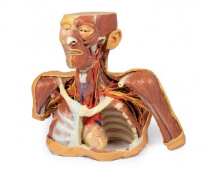

3D anatomy models - 3D head model
A human head model as an educational tool:
Anatomical models of the human head are teaching aids presenting the muscular, vascular, nervous and lymphatic structures of the facial skeleton, braincase and neck. Anatomically, the head and neck are rich in various details, often running close to each other or crossing each other. It is very important to accurately depict and visually present these structures in order to understand their functions as well as the mutual relationships between them. The above is achieved by very precisely made anatomical models, faithfully reflecting the human body. In this product category, we present anatomical models of the human head made based on radiological data and the use of modern 3D printing technology. OpenMedis' offer also includes conventional anatomical head models.
3D anatomical head models - advantages:
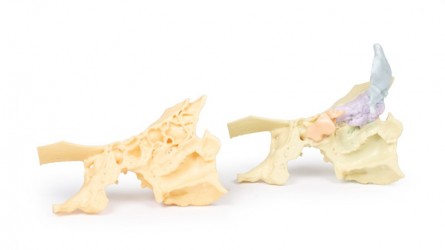
The main advantage of 3D anatomical head models is their very precise production based on CT data and 3D printing technology. These are high-fidelity anatomical models that actually reflect the dissecting preparation on the basis of which they were made. Anatomical models present anatomical structures such as muscles, nerves and vessels in their correct anatomical positions and not in an exaggerated way as in conventional models. 3D head models do not require special storage conditions and can be used in ordinary seminar rooms. Additionally, it should be noted that these products do not require disposal and their service life is very long. The undeniable advantage of 3D anatomical head models is the fact that they do not wear out over time, ensuring equal educational conditions for all students. It should also be mentioned that the colors of individual structures, adapted to the educational purposes, facilitate learning anatomy through understanding and easier remembering.
Human head models - application:
The presented anatomical models of the human head are an ideal educational tool for teaching anatomy to students of medical faculties such as medicine, physiotherapy, nursing, and emergency medical services. Currently, it is a modern solution used at many universities in Poland and abroad. 3D anatomical models of the head are perfect equipment for anatomy laboratories or an anatomy museum. They are also perfect for medical training in the field of medicine or physiotherapy, e.g. palpation anatomy courses. It is also worth mentioning the aesthetic values of anatomical head models. Taking into account the above fact, anatomical head models serve as a decorative element in teaching rooms in medical offices or constitute an element of thematic exhibitions on human anatomy.
What types of 3D anatomical models do we offer?
OpenMedis' offer includes both conventional and unconventional anatomical models depicting the human head. In this particular category, there are anatomical models of the head, presenting in detail its anatomical structure as on real dissection preparations.
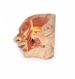
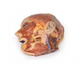
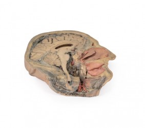
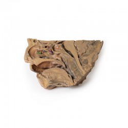
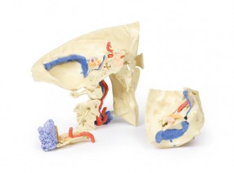
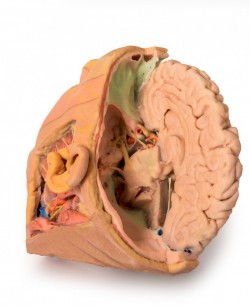
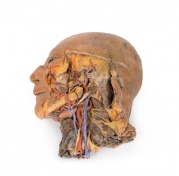
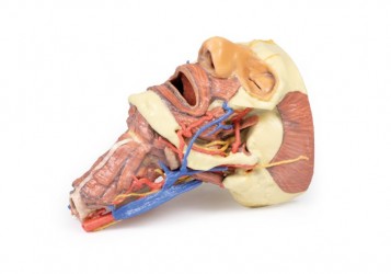
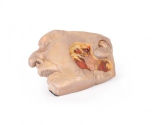
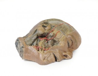
- The anatomical model of the sagittal cross-section of the head and neck (infratemporal fossa + carotid artery) is highly appreciated. This detailed head model presents anatomical structures very precisely, and the matching colors make this exhibit even more realistic, making it look like a real dissecting specimen.
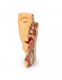
- Anatomical prints of the head, neck, shoulder and upper chest are very popular. It is an excellent teaching aid and equipment for anatomy laboratories of many medical universities.
The 3D head models in our offer are produced by a leaders in this type of solutions on the global market. Our partners are a recognizable and globally recognized manufacturer of anatomical models, medical simulators and phantoms, as well as veterinary models and simulators. It has been operating in the industry of production and distribution of teaching aids in all medical fields and veterinary medicine for over 70 years. Anatomical models from our offer are made with the utmost care and from the best quality materials. Many of them are hand-finished, which is why our anatomical models are characterized by high accuracy and care. Thanks to the use of modern 3D printing technology, it was created a unique series of 3D anatomical models. Based on radiological data, a series of models faithfully reflecting the human body were created. These advanced teaching aids are the equipment of anatomy laboratories of many Medical Universities in the World. This is a modern solution used in many countries around the world.
How can we help?
OpenMedis is an experienced manufacturer and distributor of anatomical models on the Polish market. For many years, we have been successfully equipping anatomy laboratories of medical schools and universities, medical offices, schools and museums. If you need advice on equipment for the anatomy laboratory at your institution, we will be happy to advise you on the best product range to choose. Please contact us.
We also recommend 3D anatomical models of the brain and 3D torso models, which, combined with 3D head models, will create a comprehensive set for learning the anatomy and physiology of the body.
