


Home / 3D anatomy models / 3D anatomical models of upper limb
 See our profile on Facebook
 Check our profile on Instagram
 Download a PDF file
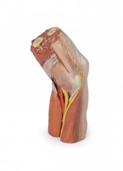

3D anatomy models - 3D anatomical models of upper limb
3D upper limb model as a teaching tool:
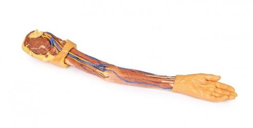
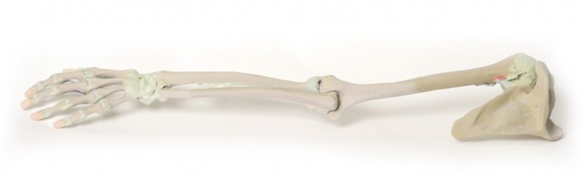
Models of the human upper limb support learning anatomy by visualizing anatomical structures such as bones, muscles, nerves, blood and lymphatic vessels. The human upper limb itself is a wealth of various anatomical details. Topographically, we can divide it into the shoulder girdle, arm, elbow, forearm and hand. From the point of view of teaching and clinical sciences, it is important to present these structures in detail in order to understand their functions and the mutual relations between them. That is why it is so important to use precisely made anatomical models in the teaching process. In this product category, we present anatomical models of the human upper limb, produced on the basis of radiological computed tomography data and using 3D printing technology. Our offer also includes conventional anatomical models of the upper limb.
3D anatomical models of the upper limb - advantages:
One of the many advantages of 3D anatomical models of the upper limb is the fact that they are made of artificial material. This means that they do not wear out over time, creating identical, equal training conditions for each student. Another undisputed advantage of 3D anatomical models of the upper limb is their extremely precise production, possible thanks to modern production technology using radiological data from CT and 3D printing. These are the so-called high-fidelity anatomical models, accurately presenting the actual dissecting preparation on the basis of which they were made. Anatomical models of the upper limb present structures such as bones, muscles, nerves, blood and lymphatic vessels in their correct anatomical courses and positions, which is particularly useful when learning topography. This is a great advantage of this type of unconventional anatomical models over conventional models that present the anatomical structure in a more exaggerated and illustrated way. Another important added value of 3D upper limb models is the fact that they do not require special storage rooms and can be used in standard teaching rooms. Additionally, it should be mentioned that these products do not require disposal and their service life is very long. Moreover, the colors of individual elements, adapted to educational purposes, facilitate learning anatomy by simplifying memorization and understanding the topography of individual structures and the mutual relations between them.
3D anatomical models of the upper limb - application:
The presented 3D models of the human upper limb are excellent educational tools that can be used in the process of teaching anatomy to students of medical faculties such as medicine, physiotherapy, nursing, emergency medical services. Currently, it is a modern solution used at many universities around the world. 3D anatomical models of the human upper limb are ideal equipment for anatomical and biological laboratories or human anatomy museums. They are also perfect for training courses in various fields of medicine. Anatomical models of the upper limb can also be used as a decorative element in teaching rooms and medical offices or constitute an element of thematic exhibitions on human anatomy.
The types of 3D anatomical models of the upper limb:
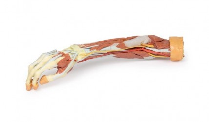
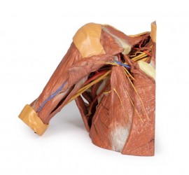
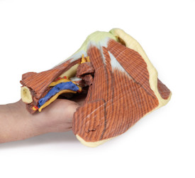
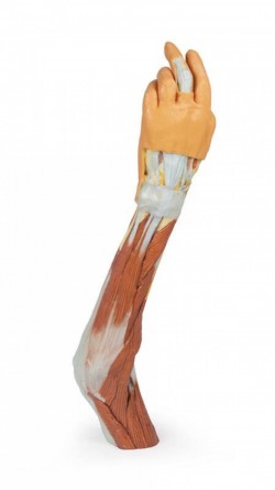
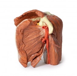
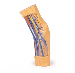
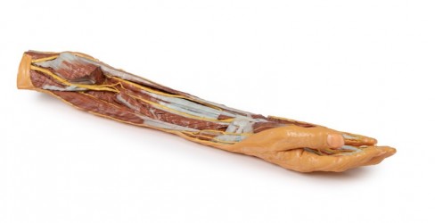
Our offer includes so-called anatomical models. conventional and unconventional human upper limbs. In this product category, we would like to present you 3D anatomical models of the upper limb, which faithfully present its anatomical structure as on real dissection preparations. There are anatomical models of the shoulder girdle, models of the entire upper limb, arm models, forearm models, and a 3D hand model.
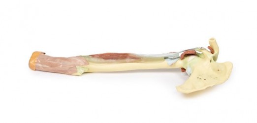
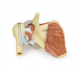
- model of the right shoulder with part of the chest
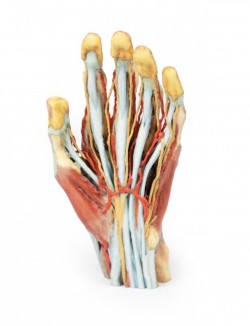
- hand model
How can we help?
OpenMedis is an experienced manufacturer and distributor of anatomical models on the Polish market. For many years, we have been successfully equipping anatomy laboratories of medical schools and universities with medical specialties, medical offices, schools and museums. If you need advice on equipment for the anatomy laboratory at your institution, we will be happy to advise you on the best selection of products. Please contact us.
We also recommend 3D anatomical models of the lower limb and 3D torso models, which, combined with 3D models of the upper limb, will create a comprehensive set for learning the anatomy and physiology of the body.
