Quality Certyficate
street: Kolejowa 2, 30-805 Cracow


Home / 3D anatomy models / 3D anatomical models of torsos
3D anatomy models
Anatomical Models
Custom tools for patient education
Veterinary simulators
Anatomical Charts
Anatomical Table 3D
Medical simulators
Medical Equipment
Type:
 See our profile on Facebook
 Check our profile on Instagram
 Download a PDF file
3D anatomy models / 3D anatomical models of torsos

This 3D printed specimen compliments our dorsal dissection specimen (AM01273) by presenting a ventral deep dissection of axial anatomy from the head, neck, axillae, thorax, and abdomen to the proximal portion of the ...
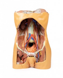
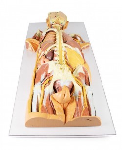
This large, multipart 3D printed specimen displays the entire male posterior abdominal wall from the diaphragm to the pelvic brim, as well as pelvic anatomy and to the proximal thigh. This same individual specimen is ...
This 3D printed specimen presents a unique view of axial anatomy, presenting a dorsal deep dissection of the head, neck, axillae, thorax, abdomen, and gluteal regions. The removal of the posterior portions of the ...
This model is a cross-section of the thorax at the level of the T6 vertebra. Beginning posteromedially at the spinal cord within the vertebral canal, then moving radially, the costovertebral joints of the 6th ribs are ...
1
3D anatomy models - 3D anatomical models of torsos
3D anatomical models of the torso as a teaching tool:
3D anatomical models of the torso are teaching aids presenting bones, muscles, internal organs, blood and lymphatic vessels, and neural structures running in the torso area. In terms of anatomy, we can distinguish the bones that make up the torso and the internal organs that are located inside it. The bones that make up the torso are: the spine, ribs, sternum and pelvic bones. The organs located inside the torso are the abdominal organs such as the liver, stomach, pancreas, small intestine and the thoracic organs such as the heart and lungs. Taking into account the division, it should be noted that the human torso is a wealth of various anatomical structures and details, often running close to each other or crossing each other. From a teaching point of view, it is important to carefully visualize these structures in order to understand their functions as well as the mutual relations between them. During the teaching process, precisely made anatomical models, faithfully presenting the human body, work great. In this product category, we present 3D anatomical models of the torso created on the basis of CT radiological data and using modern 3D printing technology. OpenMedis also offers conventional anatomical torso models.
3D anatomical models of the torso - advantages:
It should be mentioned that the 3D anatomical models of the torso are made entirely of artificial material. This means that they do not wear out over time, creating the same, equal teaching conditions for all students. An additional advantage of 3D anatomical models of the torso is their very precise production, possible thanks to the use of radiological data from CT and 3D printing technology. These are high-fidelity anatomical models, which means that they accurately reflect the actual dissecting preparation on the basis of which they were made. Anatomical torso models present structures such as bones, muscles, nerves, blood and lymphatic vessels in their correct anatomical courses and positions. This is a great advantage and superiority of this type of anatomical models compared to conventional models that present the anatomy in a more exaggerated and illustrated way. Another important added value of 3D torso models is the fact that they do not require special storage conditions and can be used in regular seminar rooms. Additionally, it should be noted that these products do not require disposal and their service life is very long. There is no doubt that the colors of individual structures, tailored to educational purposes, facilitate learning anatomy by understanding the topography of individual structures and the mutual relations between them.
3D anatomical models of the torso - application:
The presented 3D torso models are an ideal educational aid for the process of teaching anatomy to students of medical faculties such as medicine, physiotherapy, nursing, and emergency medical services. Currently, it is a modern solution used at many universities around the world. 3D anatomical models of the human torso are perfect equipment for anatomical and biological laboratories and human anatomy museums. They are also perfect for courses and training in various fields of medicine. 3D anatomical models of the torso can also serve as a decorative element in teaching rooms in medical offices or constitute an element of thematic exhibitions on human anatomy.
Types of 3D anatomical torso models:
Our offer includes so-called anatomical models conventional and unconventional torsos. In this product category, we would like to interest you in 3D anatomical models of the torso, which faithfully present its anatomical structure as on real dissection preparations. Some models show a cross-sectional view of the inside of the torso and the internal organs inside it. Our offer also includes models of the posterior body wall with innervation.
- The model of a male torso with a view of the posterior abdominal wall is very popular.
Based on radiological data, a series of models faithfully reflecting the human body were created. These advanced teaching aids are the equipment of anatomy laboratories of many Medical Universities in Poland. This is a modern solution used in many countries around the world.
How can we help?
OpenMedis is an experienced manufacturer and distributor of anatomical models on the Polish market. For many years, we have been successfully equipping anatomy laboratories of medical schools and universities, medical offices, schools and museums. If you need advice on equipment for the anatomy laboratory at your institution, we will be happy to advise you on the best product range to choose. Please contact us.
We also recommend 3D anatomical models of the head and 3D pelvic models, which, combined with 3D torso models, will create a comprehensive set for learning the anatomy and physiology of the body.
